Ito Takehito, Kimura Yasuyuki, Seki Chie, Ichise Masanori, Yokokawa Keita, Kawamura Kazunori, Takahashi Hidehiko, Higuchi Makoto, Zhang Ming-Rong, Suhara Tetsuya, Yamada Makiko
Department of Functional Brain Imaging Research, National Institute of Radiological Sciences, National Institutes for Quantum and Radiological Science and Technology, 4-9-1 Anagawa, Inage-ku, Chiba, 263-8555, Japan.
Department of Radiopharmaceuticals Development, National Institute of Radiological Sciences, National Institutes for Quantum and Radiological Science and Technology, 4-9-1 Anagawa, Inage-ku, Chiba, 263-8555, Japan.
EJNMMI Res. 2018 Jun 14;8(1):48. doi: 10.1186/s13550-018-0406-4.
The histamine H receptor is regarded as a drug target for cognitive impairments in psychiatric disorders. H receptors are expressed in neocortical areas, including the prefrontal cortex, the key region of cognitive functions such as working memory. However, the role of prefrontal H receptors in working memory has not yet been clarified. Therefore, using functional magnetic resonance imaging (fMRI) and positron emission tomography (PET) techniques, we aimed to investigate the association between the neural activity of working memory and the density of H receptors in the prefrontal cortex.
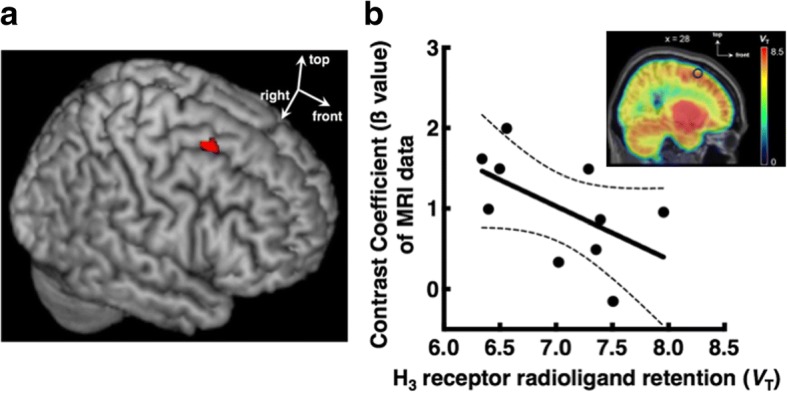
Ten healthy volunteers underwent both fMRI and PET scans. The N-back task was used to assess the neural activities related to working memory. H receptor density was measured with the selective PET radioligand [C] TASP457. The neural activity of the right dorsolateral prefrontal cortex during the performance of the N-back task was negatively correlated with the density of H receptors in this region.
Higher neural activity of working memory was associated with lower H receptor density in the right dorsolateral prefrontal cortex. This finding elucidates the role of H receptors in working memory and indicates the potential of H receptors as a therapeutic target for the cognitive impairments associated with neuropsychiatric disorders.
组胺H受体被视为精神疾病认知障碍的药物靶点。H受体在新皮质区域表达,包括前额叶皮质,而前额叶皮质是工作记忆等认知功能的关键区域。然而,前额叶H受体在工作记忆中的作用尚未阐明。因此,我们使用功能磁共振成像(fMRI)和正电子发射断层扫描(PET)技术,旨在研究工作记忆的神经活动与前额叶皮质中H受体密度之间的关联。
10名健康志愿者接受了fMRI和PET扫描。采用N-back任务评估与工作记忆相关的神经活动。使用选择性PET放射性配体[C]TASP457测量H受体密度。在执行N-back任务期间,右侧背外侧前额叶皮质的神经活动与该区域H受体密度呈负相关。
工作记忆的较高神经活动与右侧背外侧前额叶皮质中较低的H受体密度相关。这一发现阐明了H受体在工作记忆中的作用,并表明H受体作为与神经精神疾病相关的认知障碍治疗靶点的潜力。