Song Xiaoqing, Su Lining, Yin Haifeng, Dai Jin, Wei Huiping
Biology Office, Basic Medical College of Hebei North University, Zhangjiakou, Hebei 075000, P.R. China.
Exp Ther Med. 2018 Jun;15(6):5251-5260. doi: 10.3892/etm.2018.6125. Epub 2018 May 3.
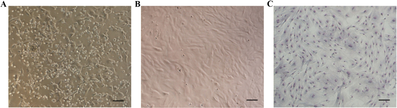
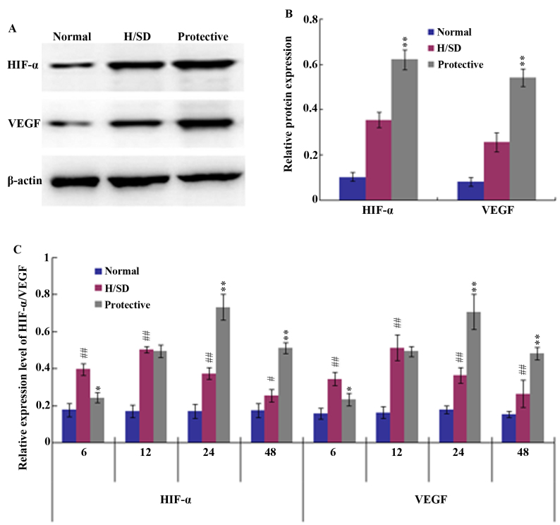
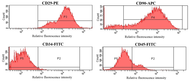
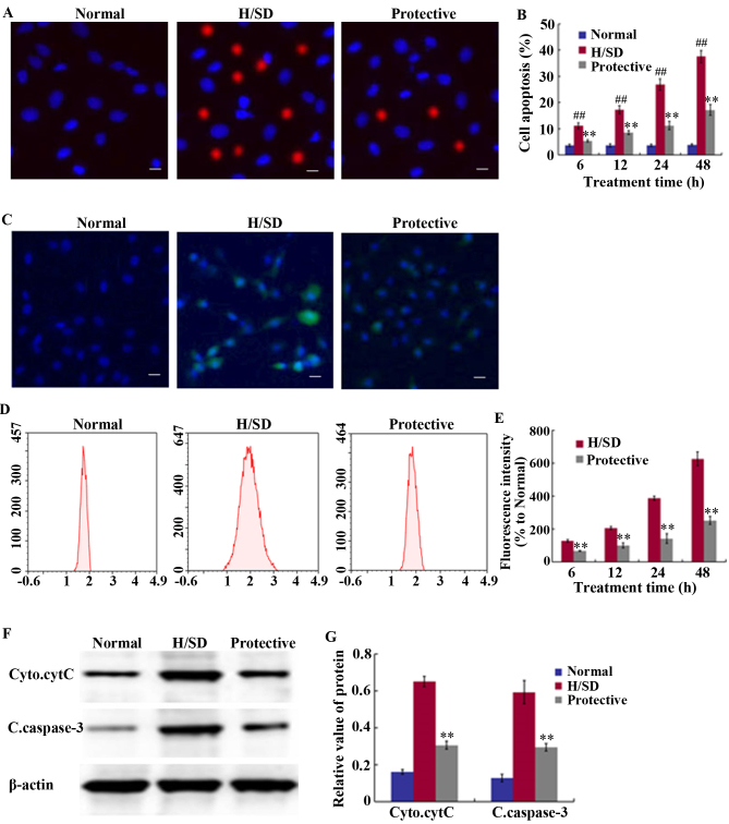
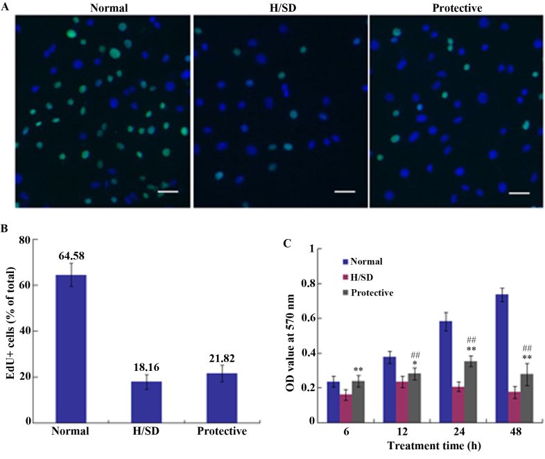
As a primary active ingredient of safflor yellow, hydroxysafflor yellow A (HSYA) exhibits notable antioxidative and neuroprotective effects. The aim of the present study was to investigate the protective effects of HSYA in mesenchymal stem cells (MSCs) exposed to hypoxia (5% O) and serum deprivation (H/SD), and to explore the mechanisms underlying HSYA-mediated protection. Under H/SD conditions, HSYA was applied to protect MSCs against injury. Cell viability, proliferation, apoptosis and reactive oxygen species (ROS) levels were determined using an 5-ethynyl-2'-deoxyuridine assay, MTT assay, Hoechst 33342/propidium iodide and 2',7'-dichlorodihydrofluorescein diacetate staining, respectively. The results revealed that 160 mg/l HSYA significantly reduced apoptosis and ROS levels compared with the H/SD group; however, HSYA demonstrated minimal effects on cell proliferation. A western blot assay demonstrated that HSYA reduced cleaved caspase-3 expression and cytC release from the mitochondria to the cytoplasm when compared with the H/SD group. In addition, western blotting and RT-qPCR analyses revealed that HSYA treatment significantly increased the expression of hypoxia inducible factor-1α (HIF-1α) and vascular endothelial growth factor (VEGF). In conclusion, the results of the current study demonstrated that HSYA exerts protective effects against H/SD-induced apoptosis in MSCs potentially via activation of the HIF-1α/VEGF signaling pathway and stabilization of the mitochondrial membrane.
作为红花黄色素的主要活性成分,羟基红花黄色素A(HSYA)具有显著的抗氧化和神经保护作用。本研究旨在探讨HSYA对暴露于低氧(5%氧气)和血清剥夺(H/SD)环境中的间充质干细胞(MSCs)的保护作用,并探究HSYA介导保护作用的潜在机制。在H/SD条件下,应用HSYA保护MSCs免受损伤。分别使用5-乙炔基-2'-脱氧尿苷检测法、MTT检测法、Hoechst 33342/碘化丙啶以及2',7'-二氯二氢荧光素二乙酸酯染色法测定细胞活力、增殖、凋亡和活性氧(ROS)水平。结果显示,与H/SD组相比,160mg/l的HSYA显著降低了细胞凋亡和ROS水平;然而,HSYA对细胞增殖的影响极小。蛋白质免疫印迹分析表明,与H/SD组相比,HSYA降低了裂解的半胱天冬酶-3的表达以及细胞色素C从线粒体向细胞质的释放。此外,蛋白质免疫印迹和RT-qPCR分析显示,HSYA处理显著增加了缺氧诱导因子-1α(HIF-1α)和血管内皮生长因子(VEGF)的表达。总之,本研究结果表明,HSYA可能通过激活HIF-1α/VEGF信号通路和稳定线粒体膜,对H/SD诱导的MSCs凋亡发挥保护作用。