Gao Bruce, Nzekwu Emeka, Cook Anthony Jonathan, Spaner Shelley Jane
The University of Calgary, Cumming School of Medicine, 3330 Hospital Dr NW, Calgary, AB, T2N 4N1, Canada.
Department of Radiology, The University of Calgary, 3330 Hospital Dr NW, Calgary, AB, T2N 4N1, Canada.
BMC Res Notes. 2018 Jun 19;11(1):396. doi: 10.1186/s13104-018-3502-7.
Nephroblastomatosis is an uncommon pathologic process characterized by the presence of persistent embryonic nephrogenic rests. Progression to Wilms tumour occurs in an estimated 35% of patients. Cure rates are based on histologic findings and disease stage and have improved from 10% in the 1920s to over 90% today.
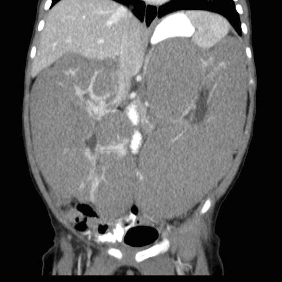
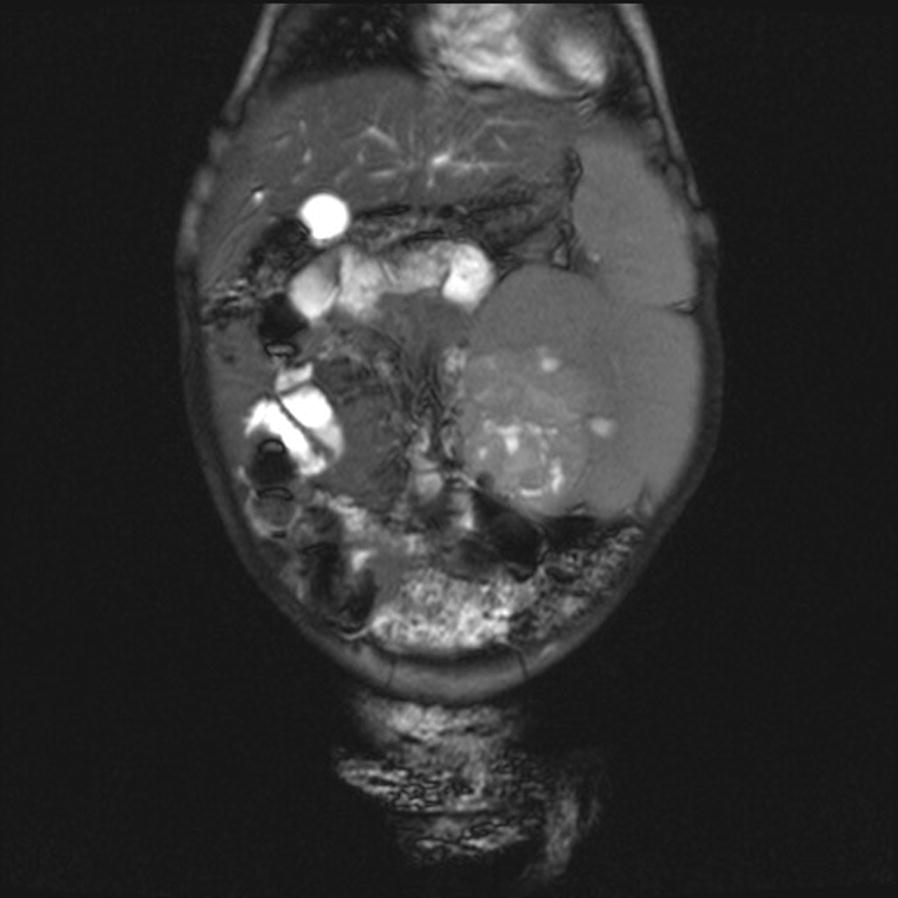
We report a case of a 9-month-old female presenting with a 2-month history of abdominal distension. Ultrasonographic and computed tomographic assessments demonstrated features consistent with bilateral, diffuse, hyperplastic perilobar nephroblastomatosis (DHPLNB) for which she underwent chemotherapy. Magnetic resonance imaging 6 weeks following commencement of chemotherapy revealed a mass concerning for unilateral Wilms tumor for which she underwent partial nephrectomy. Pathology confirmed DHPLNB with a unilateral Wilms tumor.
3.5 year radiographic follow up demonstrates complete recovery. To our knowledge, there are no similar cases with imaging depiction recently published. With potential for malignant transformation into Wilms tumour and low survival rate for late diagnosed Wilms tumors, it is important to recognize nephroblastomatosis early, both clinically and radiographically to improve overall patient prognosis.
肾母细胞瘤病是一种罕见的病理过程,其特征是存在持续性胚胎肾源性残留。估计有35%的患者会进展为肾母细胞瘤。治愈率基于组织学检查结果和疾病分期,已从20世纪20年代的10%提高到如今的90%以上。
我们报告一例9个月大的女性患者,有2个月的腹胀病史。超声和计算机断层扫描评估显示符合双侧、弥漫性、增生性叶旁肾母细胞瘤病(DHPLNB)的特征,为此她接受了化疗。化疗开始6周后的磁共振成像显示有一个疑似单侧肾母细胞瘤的肿块,为此她接受了部分肾切除术。病理证实为DHPLNB合并单侧肾母细胞瘤。
3.5年的影像学随访显示完全康复。据我们所知,最近没有类似病例的影像学描述发表。鉴于肾母细胞瘤病有恶变成为肾母细胞瘤的可能,且晚期诊断的肾母细胞瘤生存率低,早期在临床和影像学上识别肾母细胞瘤病对于改善患者总体预后很重要。