Centre for Evidence-Based Medicine, Nuffield Department of Primary Care Health Sciences, University of Oxford, Oxford OX2 6GG, UK.
University of Melbourne, Victoria, Australia.
BMJ. 2018 Jun 18;361:k2387. doi: 10.1136/bmj.k2387.
To provide an overview of the evidence on prevalence and outcomes of incidental imaging findings.
Umbrella review of systematic reviews.
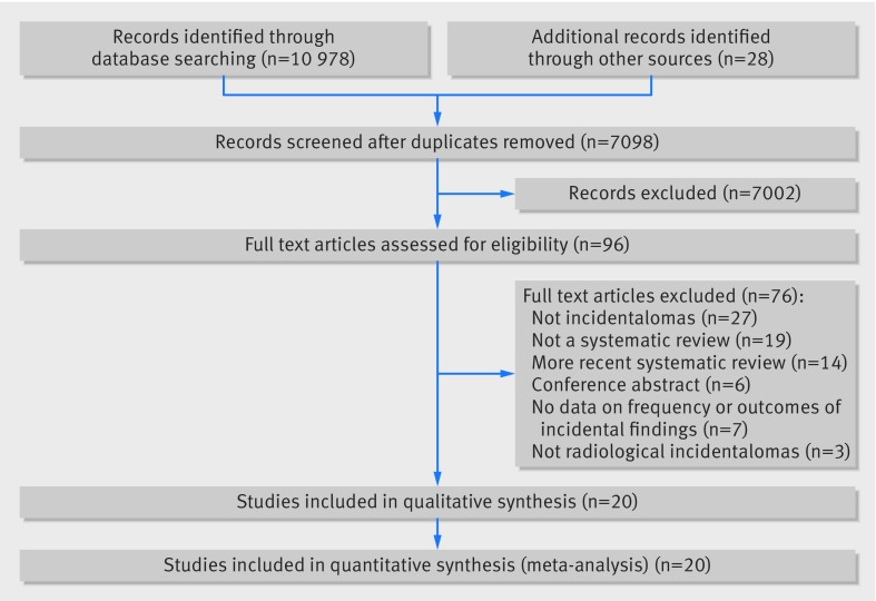
Searches of MEDLINE, EMBASE up to August 2017; screening of references in included papers.
Criteria included systematic reviews and meta-analyses of observational studies that gave a prevalence of incidental abnormalities ("incidentalomas"). An incidental imaging finding was defined as an imaging abnormality in a healthy, asymptomatic patient or an imaging abnormality in a symptomatic patient, where the abnormality was not apparently related to the patient's symptoms. Primary studies that measured the prevalence of incidentalomas in patients with a history of malignancy were also considered in sensitivity analyses.
20 systematic reviews (240 primary studies) were identified from 7098 references from the database search. Fifteen systematic reviews provided data to quantify the prevalence of incidentalomas, whereas 18 provided data to quantify the outcomes of incidentalomas (13 provided both). The prevalence of incidentalomas varied substantially between imaging tests; it was less than 5% for chest computed tomography for incidental pulmonary embolism in patients with and without cancer and whole body positron emission tomography (PET) or PET/computed tomography (for patients with and without cancer). Conversely, incidentalomas occurred in more than a third of images in cardiac magnetic resonance imaging (MRI), chest computed tomography (for incidentalomas of thorax, abdomen, spine, or heart), and computed tomography colonoscopy (for extra-colonic incidentalomas). Intermediate rates occurred with MRI of the spine (22%) and brain (22%). The rate of malignancy in incidentalomas varied substantially between organs; the prevalence of malignancy was less than 5% in incidentalomas of the brain, parotid, and adrenal gland. Extra-colonic, prostatic, and colonic incidentalomas were malignant between 10% and 20% of the time, whereas renal, thyroid, and ovarian incidentalomas were malignant around a quarter of the time. Breast incidentalomas had the highest percentage of malignancy (42%, 95% confidence interval 31% to 54%). Many assessments had high between-study heterogeneity (15 of 20 meta-analyses with I >50%).
There is large variability across different imaging techniques both in the prevalence of incidentalomas and in the prevalence of malignancy for specific organs. This umbrella review will aid clinicians and patients weigh up the pros and cons of requesting imaging scans and will help with management decisions after an incidentaloma diagnosis. Our results can underpin the creation of guidelines to assist these decisions.
PROSPERO: CRD42017075679.
提供关于偶然成像发现的患病率和结果的证据概述。
系统评价的伞式综述。
截至 2017 年 8 月,对 MEDLINE、EMBASE 的检索;纳入文献的参考文献筛选。
包括观察性研究的系统评价和荟萃分析,给出偶然异常(“偶然瘤”)的患病率。偶然成像发现被定义为健康、无症状患者的成像异常,或有症状患者的成像异常,其中异常与患者的症状显然无关。在敏感性分析中,还考虑了测量恶性肿瘤病史患者偶然瘤患病率的主要研究。
从数据库检索中,从 7098 条参考文献中确定了 20 篇系统评价(240 篇主要研究)。15 篇系统评价提供了定量偶然瘤患病率的数据,18 篇提供了偶然瘤结果的数据(13 篇同时提供了两者的数据)。成像检查之间偶然瘤的患病率差异很大;在癌症患者和非癌症患者的胸部计算机断层扫描(CT)用于偶然肺栓塞中,其患病率小于 5%,而全身正电子发射断层扫描(PET)或 PET/CT(用于癌症患者和非癌症患者)则小于 5%。相反,在心脏磁共振成像(MRI)、胸部 CT(用于偶然发现的胸部、腹部、脊柱或心脏)和 CT 结肠镜检查(用于结外偶然瘤)中,超过三分之一的图像出现偶然瘤。在脊柱(22%)和脑(22%)的 MRI 中,发生率中等。偶然瘤的恶性率在不同器官之间差异很大;脑、腮腺和肾上腺偶然瘤的恶性率小于 5%。结外、前列腺和结肠偶然瘤的恶性率为 10%至 20%,而肾、甲状腺和卵巢偶然瘤的恶性率约为四分之一。乳腺偶然瘤的恶性率最高(42%,95%置信区间 31%至 54%)。许多评估存在较大的研究间异质性(20 项荟萃分析中有 15 项 I > 50%)。
不同成像技术之间的偶然瘤患病率以及特定器官恶性肿瘤的患病率差异很大。本伞式综述将有助于临床医生和患者权衡请求成像扫描的利弊,并有助于偶然瘤诊断后的管理决策。我们的结果可以为制定指南提供依据,以协助这些决策。
PROSPERO:CRD42017075679。