• 文献检索
  • 文档翻译
  • 深度研究
  • 学术资讯
  • Suppr Zotero 插件Zotero 插件
  • 邀请有礼
  • 套餐&价格
  • 历史记录
应用&插件
Suppr Zotero 插件Zotero 插件浏览器插件Mac 客户端Windows 客户端微信小程序
定价
高级版会员购买积分包购买API积分包
服务
文献检索文档翻译深度研究API 文档MCP 服务
关于我们
关于 Suppr公司介绍联系我们用户协议隐私条款
关注我们

Suppr 超能文献

核心技术专利:CN118964589B侵权必究
粤ICP备2023148730 号-1Suppr @ 2026

文献检索

告别复杂PubMed语法,用中文像聊天一样搜索,搜遍4000万医学文献。AI智能推荐,让科研检索更轻松。

立即免费搜索

文件翻译

保留排版,准确专业,支持PDF/Word/PPT等文件格式,支持 12+语言互译。

免费翻译文档

深度研究

AI帮你快速写综述,25分钟生成高质量综述,智能提取关键信息,辅助科研写作。

立即免费体验

用于皮肤病诊断与管理的基于超声和红外的成像方式

Ultrasound and Infrared-Based Imaging Modalities for Diagnosis and Management of Cutaneous Diseases.

作者信息

Halani Sheliza, Foster F Stuart, Breslavets Maksym, Shear Neil H

机构信息

Faculty of Medicine, University of Toronto, Toronto, ON, Canada.

Medical Biophysics, Sunnybrook Health Sciences Centre, Toronto, ON, Canada.

出版信息

Front Med (Lausanne). 2018 Apr 25;5:115. doi: 10.3389/fmed.2018.00115. eCollection 2018.

DOI:10.3389/fmed.2018.00115
PMID:29922650
原文链接:https://pmc.ncbi.nlm.nih.gov/articles/PMC5996893/
Abstract

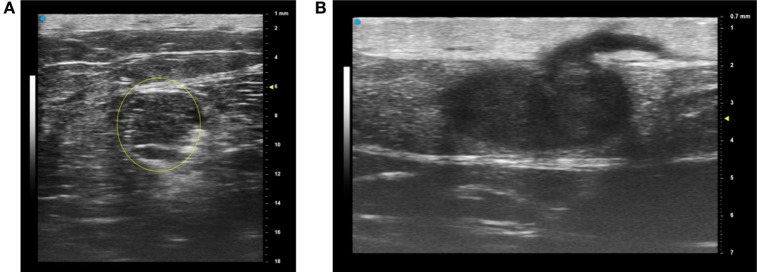
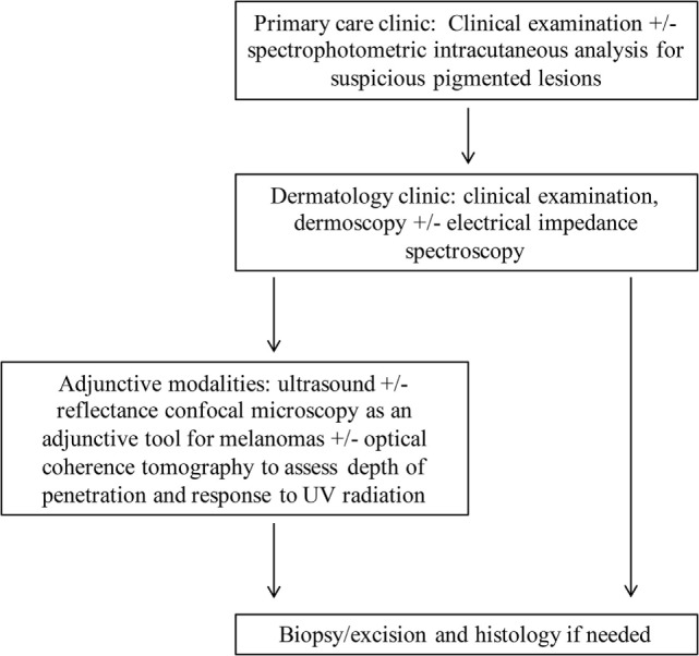
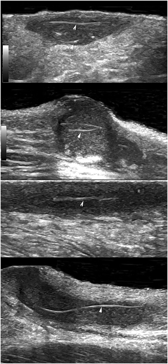
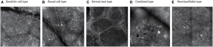
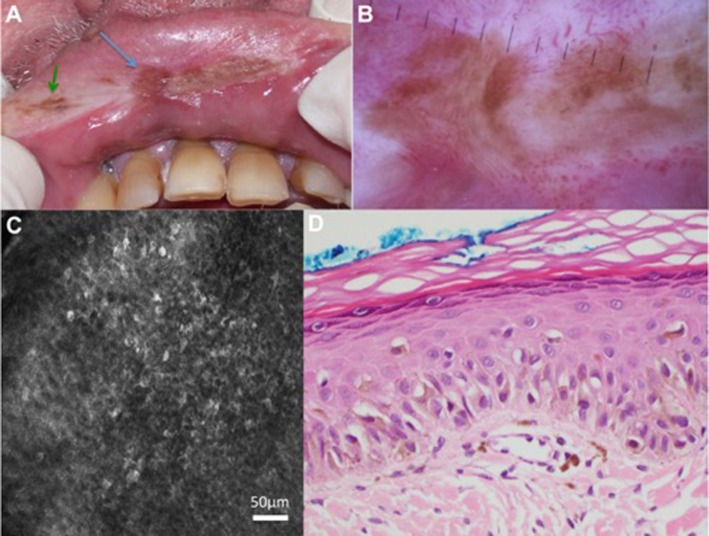
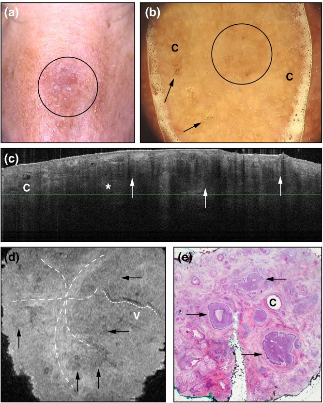
Non-invasive bedside imaging tools are becoming more prevalent for assessing cutaneous lesions. Ultrasound used at specific frequencies allows us to assess margins of lesions to minimize the extent of the biopsy that is performed and improve cosmetic outcomes. Vascularity, seen on Doppler ultrasound and contrast-enhanced ultrasound, and stiffness, assessed on tissue elastography, can help differentiate between benign and malignant lesions for clinicians to be more judicious in deciding whether to biopsy. Moreover, research has shown the efficacy in using ultrasound in monitoring flares of hidradenitis suppurativa, a disease affecting apocrine gland-rich areas of the body, for which the current gold standard involves examining and scoring inflammatory lesions with the naked eye. Infrared-based modalities have also been on the uptrend to aid in clinical decision-making regarding suspiciousness of lesions. Reflectance confocal microscopy has lateral resolution that is comparable to histopathology and it has been shown to be an appropriate adjunctive tool to dermoscopy, specifically when evaluating melanomas. Optical coherence tomography has utility in determining lesion thickness because of its depth penetration, and spectrophotometric intracutaneous analysis is becoming more popular as a tool that can be used by general practitioners to know when to refer to dermatology regarding worrisome pigmented lesions. Strides have been made to incorporate electrical impedance spectroscopy alongside dermoscopy in decision-making regarding excision, although the evidence for its use in the clincial setting remains inconclusive. This paper reviews the efficacy and drawbacks of these techniques in the field of dermatology and suggests future directions.

摘要

用于评估皮肤病变的非侵入性床边成像工具正变得越来越普遍。特定频率下使用的超声使我们能够评估病变边缘,以尽量减少活检范围并改善美容效果。通过多普勒超声和对比增强超声观察到的血管情况,以及通过组织弹性成像评估的硬度,有助于临床医生区分良性和恶性病变,从而更明智地决定是否进行活检。此外,研究表明超声在监测化脓性汗腺炎发作方面具有有效性,化脓性汗腺炎是一种影响身体富含顶泌汗腺区域的疾病,目前的金标准是用肉眼检查炎性病变并进行评分。基于红外线的方法在辅助关于病变可疑性的临床决策方面也呈上升趋势。反射式共聚焦显微镜的横向分辨率与组织病理学相当,并且已被证明是皮肤镜检查的合适辅助工具,特别是在评估黑色素瘤时。光学相干断层扫描因其深度穿透性而在确定病变厚度方面具有实用性,分光光度法皮内分析作为一种全科医生可用于判断何时将可疑色素沉着病变转诊至皮肤科的工具正变得越来越受欢迎。在关于切除的决策中,已经在将电阻抗光谱与皮肤镜检查结合使用方面取得了进展,尽管其在临床环境中使用的证据仍不明确。本文综述了这些技术在皮肤科领域的有效性和缺点,并提出了未来的发展方向。

https://cdn.ncbi.nlm.nih.gov/pmc/blobs/f8cc/5996893/351e3b1d17fc/fmed-05-00115-g006.jpg
https://cdn.ncbi.nlm.nih.gov/pmc/blobs/f8cc/5996893/b1a3efc3bee5/fmed-05-00115-g001.jpg
https://cdn.ncbi.nlm.nih.gov/pmc/blobs/f8cc/5996893/efff2a30c0ad/fmed-05-00115-g002.jpg
https://cdn.ncbi.nlm.nih.gov/pmc/blobs/f8cc/5996893/9725af1ca906/fmed-05-00115-g003.jpg
https://cdn.ncbi.nlm.nih.gov/pmc/blobs/f8cc/5996893/02d841589be3/fmed-05-00115-g004.jpg
https://cdn.ncbi.nlm.nih.gov/pmc/blobs/f8cc/5996893/6ff992822922/fmed-05-00115-g005.jpg
https://cdn.ncbi.nlm.nih.gov/pmc/blobs/f8cc/5996893/351e3b1d17fc/fmed-05-00115-g006.jpg

相似文献

1
Ultrasound and Infrared-Based Imaging Modalities for Diagnosis and Management of Cutaneous Diseases.用于皮肤病诊断与管理的基于超声和红外的成像方式
Front Med (Lausanne). 2018 Apr 25;5:115. doi: 10.3389/fmed.2018.00115. eCollection 2018.
2
Role of In Vivo Reflectance Confocal Microscopy in the Analysis of Melanocytic Lesions.体内反射共聚焦显微镜在黑素细胞性病变分析中的作用
Acta Dermatovenerol Croat. 2018 Apr;26(1):64-67.
3
Optical coherence tomography in dermatology.皮肤科中的光学相干断层扫描
G Ital Dermatol Venereol. 2015 Oct;150(5):603-15. Epub 2015 Jul 1.
4
Exploring the Utilization of Imaging Modalities in the Diagnosis of Basal Cell Carcinoma: A Scoping Review.探索成像模态在基底细胞癌诊断中的应用:一项范围综述
Cureus. 2024 Mar 12;16(3):e56047. doi: 10.7759/cureus.56047. eCollection 2024 Mar.
5
Non-invasive tools for the diagnosis of cutaneous melanoma.用于诊断皮肤黑色素瘤的非侵入性工具。
Skin Res Technol. 2017 Aug;23(3):261-271. doi: 10.1111/srt.12350. Epub 2016 Nov 22.
6
Differentiation Between Benign and Malignant Pigmented Skin Tumours Using Bedside Diagnostic Imaging Technologies: A Pilot Study.利用床边诊断成像技术鉴别良恶性色素性皮肤肿瘤:一项初步研究。
Acta Derm Venereol. 2022 Jan 26;102:adv00634. doi: 10.2340/actadv.v101.571.
7
Current uses of diagnostic high-frequency US in dermatology.诊断性高频超声在皮肤科的当前应用。
Eur J Radiol. 1998 May;27 Suppl 2:S215-23. doi: 10.1016/s0720-048x(98)00065-5.
8
Noninvasive diagnosis in dermatology.皮肤科的非侵入性诊断。
J Dtsch Dermatol Ges. 2017 Oct;15(10):999-1016. doi: 10.1111/ddg.13347.
9
Dermoscopy, Confocal Microscopy and other Non-invasive Tools for the Diagnosis of Non-Melanoma Skin Cancers and Other Skin Conditions.皮肤镜检查、共聚焦显微镜检查及其他用于诊断非黑色素瘤皮肤癌和其他皮肤疾病的非侵入性工具。
Acta Derm Venereol. 2017;Suppl 218:22-30. doi: 10.2340/00015555-2720. Epub 2017 Jul 5.
10
Role of modern imaging techniques for the diagnosis of lichen planus.现代成像技术在扁平苔藓诊断中的作用
Exp Ther Med. 2019 Feb;17(2):1052-1060. doi: 10.3892/etm.2018.6974. Epub 2018 Nov 16.

引用本文的文献

1
The Role of FDG-PET in the Evaluation of Hidradenitis Suppurativa: A Systematic Review.18F-氟脱氧葡萄糖正电子发射断层扫描在化脓性汗腺炎评估中的作用:一项系统评价
J Clin Med. 2023 Aug 24;12(17):5491. doi: 10.3390/jcm12175491.
2
1.7-Micron Optical Coherence Tomography Angiography for Characterization of Skin Lesions-A Feasibility Study.1.7 微米光学相干断层扫描血管造影术用于皮肤病变的特征分析:一项可行性研究。
IEEE Trans Med Imaging. 2021 Sep;40(9):2507-2512. doi: 10.1109/TMI.2021.3081066. Epub 2021 Aug 31.
3
Non-contrast-enhanced 3-Tesla Magnetic Resonance Imaging Using Surface-coil and Sonography for Assessment of Hidradenitis Suppurativa Lesions.

本文引用的文献

1
The validity and reliability of using ultrasound elastography to measure cutaneous stiffness, a systematic review.使用超声弹性成像测量皮肤硬度的有效性和可靠性:一项系统综述
Int J Burns Trauma. 2017 Dec 20;7(7):124-141. eCollection 2017.
2
Noninvasive diagnosis in dermatology.皮肤科的非侵入性诊断。
J Dtsch Dermatol Ges. 2017 Oct;15(10):999-1016. doi: 10.1111/ddg.13347.
3
Ultrasound Evaluation as a Complementary Test in Hidradenitis Suppurativa: Proposal of a Standarized Report.超声评估作为化脓性汗腺炎的补充检查:标准化报告建议
非增强 3.0T 磁共振成像联合表面线圈及超声评估化脓性汗腺炎皮损。
Acta Derm Venereol. 2020 Nov 12;100(18):adv00317. doi: 10.2340/00015555-3639.
4
A Scoping Review of Non-invasive Imaging Modalities in Dermatological Disease: Potential Novel Biomarkers in Hidradenitis Suppurativa.皮肤病无创成像模式的范围综述:化脓性汗腺炎潜在的新型生物标志物
Front Med (Lausanne). 2019 Nov 6;6:253. doi: 10.3389/fmed.2019.00253. eCollection 2019.
5
Utility of a gel stand-off pad in the detection of Doppler signal on focal nodular lesions of the skin.凝胶隔离垫在皮肤局灶性结节病变多普勒信号检测中的应用。
J Ultrasound. 2020 Mar;23(1):45-53. doi: 10.1007/s40477-019-00376-3. Epub 2019 Mar 29.
Dermatol Surg. 2017 Aug;43(8):1065-1073. doi: 10.1097/DSS.0000000000001147.
4
Analysis of an electrical impedance spectroscopy system in short-term digital dermoscopy imaging of melanocytic lesions.分析电阻抗光谱系统在黑素细胞病变短期数字皮肤镜成像中的应用。
Br J Dermatol. 2017 Nov;177(5):1432-1438. doi: 10.1111/bjd.15595. Epub 2017 Oct 11.
5
Imaging Blood Vessel Morphology in Skin: Dynamic Optical Coherence Tomography as a Novel Potential Diagnostic Tool in Dermatology.皮肤血管形态成像:动态光学相干断层扫描作为皮肤病学中一种新型潜在诊断工具
Dermatol Ther (Heidelb). 2017 Jun;7(2):187-202. doi: 10.1007/s13555-017-0175-4. Epub 2017 Mar 3.
6
Dermatologist-level classification of skin cancer with deep neural networks.基于深度神经网络的皮肤癌皮肤科医生级分类。
Nature. 2017 Feb 2;542(7639):115-118. doi: 10.1038/nature21056. Epub 2017 Jan 25.
7
Comparative study of presurgical skin infiltration depth measurements of melanocytic lesions with OCT and high frequency ultrasound.光学相干断层扫描(OCT)与高频超声用于黑素细胞性病变术前皮肤浸润深度测量的对比研究
J Biophotonics. 2017 Jun;10(6-7):854-861. doi: 10.1002/jbio.201600139. Epub 2016 Dec 23.
8
Non-invasive tools for the diagnosis of cutaneous melanoma.用于诊断皮肤黑色素瘤的非侵入性工具。
Skin Res Technol. 2017 Aug;23(3):261-271. doi: 10.1111/srt.12350. Epub 2016 Nov 22.
9
Evaluation of electrical impedance spectroscopy as an adjunct to dermoscopy in short-term monitoring of atypical melanocytic lesions.评估电阻抗光谱法作为皮肤镜辅助手段在非典型黑素细胞性病变短期监测中的应用。
Dermatol Pract Concept. 2016 Oct 31;6(4):1-6. doi: 10.5826/dpc.0604a01. eCollection 2016 Oct.
10
Analysis of debrided and non-debrided invasive squamous cell carcinoma skin lesions by in vivo reflectance confocal microscopy before and after therapy.治疗前后通过体内反射共聚焦显微镜对清创和未清创的浸润性鳞状细胞癌皮肤病变进行分析。
Lasers Med Sci. 2017 Jan;32(1):211-219. doi: 10.1007/s10103-016-2104-7. Epub 2016 Nov 12.