• 文献检索
  • 文档翻译
  • 深度研究
  • 学术资讯
  • Suppr Zotero 插件Zotero 插件
  • 邀请有礼
  • 套餐&价格
  • 历史记录
应用&插件
Suppr Zotero 插件Zotero 插件浏览器插件Mac 客户端Windows 客户端微信小程序
定价
高级版会员购买积分包购买API积分包
服务
文献检索文档翻译深度研究API 文档MCP 服务
关于我们
关于 Suppr公司介绍联系我们用户协议隐私条款
关注我们

Suppr 超能文献

核心技术专利:CN118964589B侵权必究
粤ICP备2023148730 号-1Suppr @ 2026

文献检索

告别复杂PubMed语法,用中文像聊天一样搜索,搜遍4000万医学文献。AI智能推荐,让科研检索更轻松。

立即免费搜索

文件翻译

保留排版,准确专业,支持PDF/Word/PPT等文件格式,支持 12+语言互译。

免费翻译文档

深度研究

AI帮你快速写综述,25分钟生成高质量综述,智能提取关键信息,辅助科研写作。

立即免费体验

光学相干断层扫描结果作为接受雷珠单抗治疗的视网膜分支静脉阻塞患者临床病程的预测指标。

Optical coherence tomography findings as a predictor of clinical course in patients with branch retinal vein occlusion treated with ranibizumab.

机构信息

Department of Ophthalmology, St. Marianna University School of Medicine, Kawasaki, Kanagawa, Japan.

出版信息

PLoS One. 2018 Jun 20;13(6):e0199552. doi: 10.1371/journal.pone.0199552. eCollection 2018.

DOI:10.1371/journal.pone.0199552
PMID:29924853
原文链接:https://pmc.ncbi.nlm.nih.gov/articles/PMC6010278/
Abstract

PURPOSE

To examine the relationship between optical coherence tomography (OCT) images and clinical course in eyes with branch retinal vein occlusion (BRVO) treated with intravitreal ranibizumab injection (IVR).

DESIGN

Prospective cohort study.

PARTICIPANTS

Thirty eyes of 30 patients with BRVO treated with IVR.

METHODS

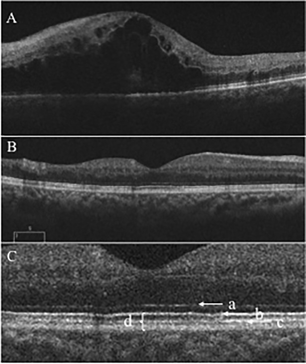
All patients received 1 initial IVR followed by repeated injections in the pro re nata (PRN) regimen. Correlations between logarithm of minimum angle of resolution best-corrected visual acuity (logMAR BCVA) or number of IVRs after 12 months and OCT parameters including the external limiting membrane (ELM), ellipsoid zone (EZ), interdigitation zone (IZ), and photoreceptor outer segment (PROS) length at first resolution of macular edema (ME) were assessed. Resolution of ME was defined as central foveal thickness <300 μm and the absence of subretinal fluid. OCT parameters influencing BCVA and number of IVRs were evaluated using multivariate analysis. Correlations between nonperfusion areas (NPAs) and thinning areas and changes in retinal thickness of BRVO-affected areas were assessed.

RESULTS

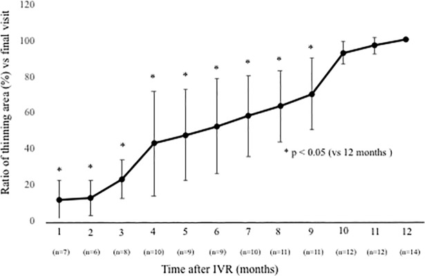
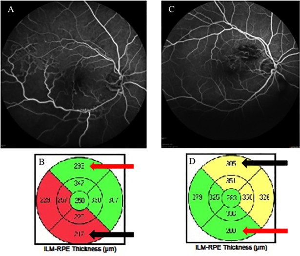
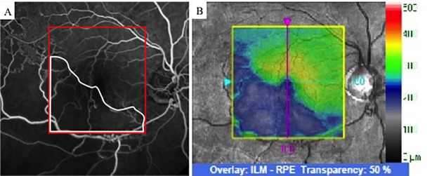
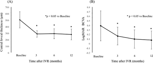
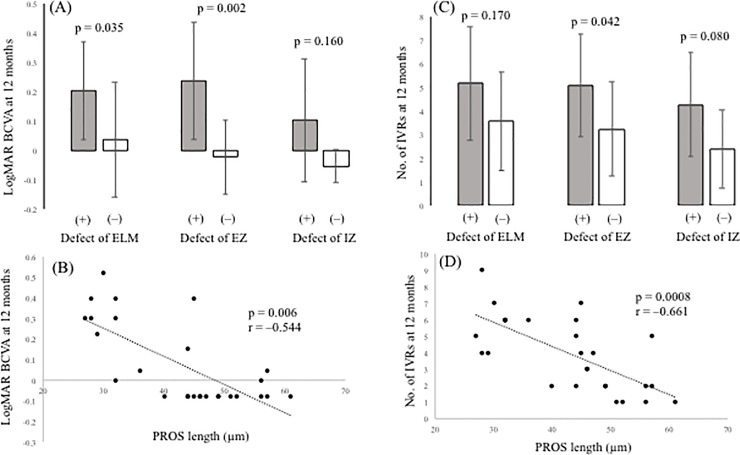
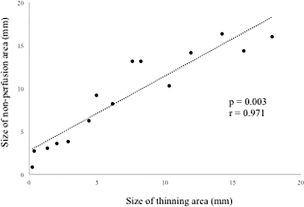
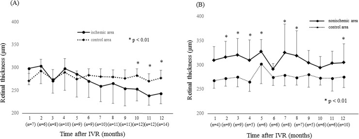
Of the 30 patients, 27 completed this study and were included in the statistical analyses. The mean logMAR BCVA at 3, 6, and 12 months was 0.16 ± 0.19, 0.09 ± 0.20, and 0.07 ± 0.20, respectively, which improved significantly from baseline at each visit (p < 0.0001, respectively), while the mean number of IVRs at 12 months was 3.9 ± 2.2. The mean number of IVRs for the first resolution of ME was 1.6 ± 0.8. Eyes with ELM and EZ defects at the points of first resolution of ME were correlated with a significantly lower BCVA at 12 months compared with eyes with preserved ELMs and EZs (p = 0.035, p = 0.002, respectively). However, eyes with IZ defects at the points of first resolution of ME were not correlated with a significantly lower BCVA at 12 months compared with eyes with preserved IZs (p = 0.160). Defects in the EZ at the points of first resolution of ME significantly affected the number of IVRs at 12 months (p = 0.042), although the ELM and IZ did not. PROS length at the points of first resolution of ME was significantly correlated with BCVA and number of IVRs at 12 months (p = 0.006, p = 0.0008, respectively). In multivariate analysis, PROS length at the points of first resolution of ME had the most significant effect on BCVA and number of IVRs (p = 0.013, p = 0.012, respectively). NPA size on fluorescein angiography and thinning area on OCT within the macular area showed a significant correlation (p = 0.003, r = 0.971). The retinal thickness of ischemic BRVO-affected areas was significantly less than that of control areas at 10, 11, and 12 months (p = 0.001, p = 0.005, p = 0.003, respectively).

CONCLUSION

We showed that the 1+PRN regimen may be a useful therapy for ME due to BRVO. In addition, PROS length at points of first resolution of ME appears to be a good indicator of BCVA and number of IVRs in BRVO patients.

摘要

目的

研究玻璃体内注射雷珠单抗(IVR)治疗视网膜分支静脉阻塞(BRVO)后 OCT 图像与临床病程的关系。

设计

前瞻性队列研究。

参与者

30 例 BRVO 患者的 30 只眼接受 IVR 治疗。

方法

所有患者均接受 1 次初始 IVR,随后根据需要进行重复注射(PRN)。评估黄斑水肿(ME)首次消退时的对数最小角分辨率最佳矫正视力(logMAR BCVA)或 12 个月后 IVR 次数与 OCT 参数(包括外节膜(ELM)、椭圆体带(EZ)、交错区(IZ)和光感受器外节(PROS)长度)之间的相关性。ME 消退的定义为中央视网膜厚度<300μm且无脉络膜下积液。使用多元分析评估影响 BCVA 和 IVR 次数的 OCT 参数。评估荧光素血管造影中的无灌注区(NPA)大小和 OCT 内黄斑区的变薄区与 BRVO 受累区视网膜厚度变化之间的相关性。

结果

30 例患者中,27 例完成了本研究并纳入了统计分析。在第 3、6 和 12 个月时,平均 logMAR BCVA 分别为 0.16±0.19、0.09±0.20 和 0.07±0.20,与基线相比,在每次就诊时均显著提高(分别为 p<0.0001),而 12 个月时平均 IVR 次数为 3.9±2.2。ME 首次消退时的平均 IVR 次数为 1.6±0.8。ME 首次消退时存在 ELM 和 EZ 缺陷的眼,与 12 个月时 BCVA 显著降低相关(p=0.035,p=0.002)。然而,ME 首次消退时存在 IZ 缺陷的眼,与 12 个月时 BCVA 降低不显著相关(p=0.160)。ME 首次消退时 EZ 缺陷显著影响 12 个月时的 IVR 次数(p=0.042),而 ELM 和 IZ 则无显著影响。ME 首次消退时 PROS 长度与 12 个月时的 BCVA 和 IVR 次数显著相关(p=0.006,p=0.0008)。多元分析显示,ME 首次消退时的 PROS 长度对 BCVA 和 IVR 次数的影响最大(p=0.013,p=0.012)。荧光素血管造影中的 NPA 大小和 OCT 内黄斑区的变薄区之间存在显著相关性(p=0.003,r=0.971)。缺血性 BRVO 受累区的视网膜厚度在 10、11 和 12 个月时均显著低于对照组(p=0.001,p=0.005,p=0.003)。

结论

我们表明,1+PRN 方案可能是治疗 BRVO 引起的 ME 的有效治疗方法。此外,ME 首次消退时 PROS 长度似乎是 BRVO 患者 BCVA 和 IVR 次数的良好指标。

https://cdn.ncbi.nlm.nih.gov/pmc/blobs/53ef/6010278/704b8bb39cb2/pone.0199552.g009.jpg
https://cdn.ncbi.nlm.nih.gov/pmc/blobs/53ef/6010278/c8981ba8a629/pone.0199552.g001.jpg
https://cdn.ncbi.nlm.nih.gov/pmc/blobs/53ef/6010278/604f01d673b4/pone.0199552.g002.jpg
https://cdn.ncbi.nlm.nih.gov/pmc/blobs/53ef/6010278/1a99ce6a4251/pone.0199552.g003.jpg
https://cdn.ncbi.nlm.nih.gov/pmc/blobs/53ef/6010278/4ce09707be76/pone.0199552.g004.jpg
https://cdn.ncbi.nlm.nih.gov/pmc/blobs/53ef/6010278/b66ebf850e9c/pone.0199552.g005.jpg
https://cdn.ncbi.nlm.nih.gov/pmc/blobs/53ef/6010278/88f372e1aa5d/pone.0199552.g006.jpg
https://cdn.ncbi.nlm.nih.gov/pmc/blobs/53ef/6010278/bd73ab8621cf/pone.0199552.g007.jpg
https://cdn.ncbi.nlm.nih.gov/pmc/blobs/53ef/6010278/04f91513f6c7/pone.0199552.g008.jpg
https://cdn.ncbi.nlm.nih.gov/pmc/blobs/53ef/6010278/704b8bb39cb2/pone.0199552.g009.jpg

相似文献

1
Optical coherence tomography findings as a predictor of clinical course in patients with branch retinal vein occlusion treated with ranibizumab.光学相干断层扫描结果作为接受雷珠单抗治疗的视网膜分支静脉阻塞患者临床病程的预测指标。
PLoS One. 2018 Jun 20;13(6):e0199552. doi: 10.1371/journal.pone.0199552. eCollection 2018.
2
Macular vessel reduction as predictor for recurrence of macular oedema requiring repeat intravitreal ranibizumab injection in eyes with branch retinal vein occlusion.脉络膜血管减少可预测分支型视网膜静脉阻塞患者黄斑水肿复发,需再次玻璃体内注射雷珠单抗。
Br J Ophthalmol. 2019 Oct;103(10):1367-1372. doi: 10.1136/bjophthalmol-2018-312769. Epub 2018 Dec 4.
3
Intravitreal ranibizumab for macular edema secondary to retinal vein occlusion.玻璃体内雷珠单抗治疗视网膜静脉阻塞所致黄斑水肿。
Ophthalmologica. 2012;227(3):132-8. doi: 10.1159/000334906. Epub 2011 Dec 29.
4
Multimodal imaging to distinguish microvascular and morphological changes in retinal vein occlusion after intravitreal ranibizumab with or without triamcinolone acetonide injection.多模态成像在玻璃体内雷珠单抗联合或不联合曲安奈德注射治疗视网膜静脉阻塞后的微血管和形态学改变中的鉴别诊断。
BMC Ophthalmol. 2024 Aug 20;24(1):354. doi: 10.1186/s12886-024-03619-y.
5
Comparison of one and three initial monthly intravitreal ranibizumab injection in patients with macular edema secondary to branch retinal vein occlusion.视网膜分支静脉阻塞继发黄斑水肿患者初始每月一次与每月三次玻璃体内注射雷珠单抗的比较
Int J Ophthalmol. 2018 Sep 18;11(9):1534-1538. doi: 10.18240/ijo.2018.09.17. eCollection 2018.
6
Twenty-Four Month Results of Intravitreal Ranibizumab for Macular Edema after Branch Retinal Vein Occlusion: Visual Outcomes and Resolution of Macular Edema.玻璃体内雷珠单抗治疗分支型视网膜静脉阻塞继发黄斑水肿的 24 个月疗效:视力结果和黄斑水肿消退情况。
Semin Ophthalmol. 2021 Oct 3;36(7):482-489. doi: 10.1080/08820538.2021.1890147. Epub 2021 Feb 22.
7
Spectral-Domain OCT Predictors of Visual Outcomes after Ranibizumab Treatment for Macular Edema Resulting from Retinal Vein Occlusion.雷珠单抗治疗视网膜静脉阻塞所致黄斑水肿后视力预后的频域光学相干断层扫描预测指标
Ophthalmol Retina. 2020 Jan;4(1):67-76. doi: 10.1016/j.oret.2019.08.009. Epub 2019 Aug 28.
8
Macular function following intravitreal ranibizumab for macular edema associated with branch retinal vein occlusion.玻璃体内注射雷珠单抗治疗视网膜分支静脉阻塞相关黄斑水肿后的黄斑功能
Doc Ophthalmol. 2019 Aug;139(1):67-74. doi: 10.1007/s10633-019-09696-5. Epub 2019 Apr 12.
9
Comparing ranibizumab, dexamethasone implant, and combined therapy for macular edema secondary to branch retinal vein occlusion: a clinical trial.比较雷珠单抗、地塞米松植入物和联合治疗对视网膜分支静脉阻塞引起的黄斑水肿:一项临床试验。
Int Ophthalmol. 2024 Jun 24;44(1):262. doi: 10.1007/s10792-024-03158-x.
10
Individualized Stabilization Criteria-Driven Ranibizumab versus Laser in Branch Retinal Vein Occlusion: Six-Month Results of BRIGHTER.基于个体化稳定标准的雷珠单抗与激光治疗分支静脉阻塞:BRIGHTER 研究 6 个月结果。
Ophthalmology. 2016 Jun;123(6):1332-44. doi: 10.1016/j.ophtha.2016.02.030. Epub 2016 Mar 30.

引用本文的文献

1
Predictors of macular edema recurrence and visual outcomes in branch retinal vein occlusion treated with aflibercept or ranibizumab.阿柏西普或雷珠单抗治疗视网膜分支静脉阻塞后黄斑水肿复发及视力预后的预测因素
Int Ophthalmol. 2025 Mar 14;45(1):96. doi: 10.1007/s10792-025-03480-y.
2
Impact of anti-VEGF therapy on distinctive retina layers in patients with macular edema secondary to branch retinal vein occlusion.抗 VEGF 治疗对分支视网膜静脉阻塞继发黄斑水肿患者不同视网膜层的影响。
BMC Ophthalmol. 2023 May 25;23(1):235. doi: 10.1186/s12886-023-02981-7.
3
Visual Prognostic Factors in Eyes with Subretinal Fluid Associated with Branch Retinal Vein Occlusion.

本文引用的文献

1
Outcome of "treat and monitor" regimen of aflibercept and ranibizumab in macular edema secondary to non-ischemic branch retinal vein occlusion.阿柏西普和雷珠单抗“治疗并监测”方案用于非缺血性视网膜分支静脉阻塞继发黄斑水肿的疗效
Int Ophthalmol. 2019 Jan;39(1):145-153. doi: 10.1007/s10792-017-0798-6. Epub 2017 Dec 22.
2
Foveal Microstructure Analysis in Eyes with Diabetic Macular Edema Treated with Vitrectomy.糖尿病性黄斑水肿患者玻璃体切除术后的中心凹微观结构分析。
Adv Ther. 2017 Sep;34(9):2139-2149. doi: 10.1007/s12325-017-0598-4. Epub 2017 Aug 14.
3
Efficacy of intravitreal bevacizumab for macular edema following branch retinal vein occlusion stratified by baseline visual acuity.
视网膜分支静脉阻塞相关的视网膜下液眼中的视觉预后因素
J Clin Med. 2023 Apr 17;12(8):2909. doi: 10.3390/jcm12082909.
4
Short-Term Hyperuricemia Leads to Structural Retinal Changes That Can be Reversed by Serum Uric Acid Lowering Agents in Mice.短期高尿酸血症导致的视网膜结构变化可被小鼠血清尿酸降低剂逆转。
Invest Ophthalmol Vis Sci. 2022 Sep 1;63(10):8. doi: 10.1167/iovs.63.10.8.
5
Which Explanatory Variables Contribute to the Classification of Good Visual Acuity over Time in Patients with Branch Retinal Vein Occlusion with Macular Edema Using Machine Learning?使用机器学习时,哪些解释变量有助于对伴有黄斑水肿的视网膜分支静脉阻塞患者的良好视力随时间进行分类?
J Clin Med. 2022 Jul 4;11(13):3903. doi: 10.3390/jcm11133903.
6
Foveal Intraretinal Fluid Localization Affects the Visual Prognosis of Branch Retinal Vein Occlusion.黄斑区视网膜内液的定位影响视网膜分支静脉阻塞的视觉预后。
J Clin Med. 2022 Jun 20;11(12):3540. doi: 10.3390/jcm11123540.
7
Comparison of Choroidal Thickness, Foveal Avascular Zone, and Macular Capillary Density in Macular Edema Secondary to Branch Retinal Vein Occlusion Treated with Ranibizumab or Aflibercept-A Prospective Study.比较雷珠单抗和阿柏西普治疗分支型视网膜静脉阻塞继发黄斑水肿的脉络膜厚度、中心无血管区和黄斑毛细血管密度:一项前瞻性研究。
Medicina (Kaunas). 2022 Apr 14;58(4):540. doi: 10.3390/medicina58040540.
8
Ellipsoid Zone Defects in Retinal Vein Occlusion Correlates With Visual Acuity Prognosis: SCORE2 Report 14.视网膜静脉阻塞的椭圆体带缺损与视力预后相关:SCORE2 报告 14 期。
Transl Vis Sci Technol. 2021 Mar 1;10(3):31. doi: 10.1167/tvst.10.3.31.
9
Ocular microcirculation changes, measured with laser speckle flowgraphy and optical coherence tomography angiography, in branch retinal vein occlusion with macular edema treated by ranibizumab.经雷射散斑血流图和光相干断层扫描血管造影术测量,接受雷珠单抗治疗的视网膜分支静脉阻塞伴黄斑水肿的眼微循环变化。
Int Ophthalmol. 2021 Jan;41(1):151-162. doi: 10.1007/s10792-020-01562-7. Epub 2020 Sep 7.
10
Macular Pigment Optical Density and Photoreceptor Outer Segment Length as Predisease Biomarkers for Age-Related Macular Degeneration.黄斑色素光密度和光感受器外段长度作为年龄相关性黄斑变性的疾病前生物标志物
J Clin Med. 2020 May 5;9(5):1347. doi: 10.3390/jcm9051347.
玻璃体内注射贝伐单抗治疗视网膜分支静脉阻塞后黄斑水肿的疗效,按基线视力分层分析
Graefes Arch Clin Exp Ophthalmol. 2017 Apr;255(4):691-697. doi: 10.1007/s00417-016-3535-3. Epub 2016 Oct 29.
4
Macular Inner Retinal Layer Thickening and Outer Retinal Layer Damage Correlate With Visual Acuity During Remission in Behcet's Disease.白塞病缓解期黄斑视网膜内层增厚和外层视网膜损伤与视力相关。
Invest Ophthalmol Vis Sci. 2016 Oct 1;57(13):5470-5478. doi: 10.1167/iovs.16-19568.
5
Automated Quantification of Nonperfusion in Three Retinal Plexuses Using Projection-Resolved Optical Coherence Tomography Angiography in Diabetic Retinopathy.使用投影分辨光学相干断层扫描血管造影术对糖尿病视网膜病变中三个视网膜神经丛的无灌注进行自动定量分析。
Invest Ophthalmol Vis Sci. 2016 Oct 1;57(13):5101-5106. doi: 10.1167/iovs.16-19776.
6
RANIBIZUMAB FOR MACULAR EDEMA AFTER BRANCH RETINAL VEIN OCCLUSION: One Initial Injection Versus Three Monthly Injections.雷珠单抗治疗视网膜分支静脉阻塞后黄斑水肿:单次初始注射与每月三次注射的比较
Retina. 2017 Apr;37(4):702-709. doi: 10.1097/IAE.0000000000001224.
7
Wide-field optical coherence tomography angiography using extended field imaging technique to evaluate the nonperfusion area in retinal vein occlusion.使用扩展视野成像技术的广角光学相干断层扫描血管造影术评估视网膜静脉阻塞中的无灌注区。
Clin Ophthalmol. 2016 Jul 13;10:1291-5. doi: 10.2147/OPTH.S108630. eCollection 2016.
8
Predicting outcomes to anti-vascular endothelial growth factor (VEGF) therapy in diabetic macular oedema: a review of the literature.预测糖尿病性黄斑水肿抗血管内皮生长因子(VEGF)治疗的结果:文献综述
Br J Ophthalmol. 2016 Dec;100(12):1596-1604. doi: 10.1136/bjophthalmol-2016-308388. Epub 2016 May 26.
9
Automated Quantification of Capillary Nonperfusion Using Optical Coherence Tomography Angiography in Diabetic Retinopathy.利用光学相干断层扫描血管造影术自动定量分析糖尿病视网膜病变中的毛细血管无灌注情况。
JAMA Ophthalmol. 2016 Apr;134(4):367-73. doi: 10.1001/jamaophthalmol.2015.5658.
10
Prediction of Retinal Ischemia in Branch Retinal Vein Occlusion: Spectral-Domain Optical Coherence Tomography Study.视网膜分支静脉阻塞中视网膜缺血的预测:频域光学相干断层扫描研究
Invest Ophthalmol Vis Sci. 2015 Oct;56(11):6622-9. doi: 10.1167/iovs.15-17678.