• 文献检索
  • 文档翻译
  • 深度研究
  • 学术资讯
  • Suppr Zotero 插件Zotero 插件
  • 邀请有礼
  • 套餐&价格
  • 历史记录
应用&插件
Suppr Zotero 插件Zotero 插件浏览器插件Mac 客户端Windows 客户端微信小程序
定价
高级版会员购买积分包购买API积分包
服务
文献检索文档翻译深度研究API 文档MCP 服务
关于我们
关于 Suppr公司介绍联系我们用户协议隐私条款
关注我们

Suppr 超能文献

核心技术专利:CN118964589B侵权必究
粤ICP备2023148730 号-1Suppr @ 2026

文献检索

告别复杂PubMed语法,用中文像聊天一样搜索,搜遍4000万医学文献。AI智能推荐,让科研检索更轻松。

立即免费搜索

文件翻译

保留排版,准确专业,支持PDF/Word/PPT等文件格式,支持 12+语言互译。

免费翻译文档

深度研究

AI帮你快速写综述,25分钟生成高质量综述,智能提取关键信息,辅助科研写作。

立即免费体验

颅神经孔:第二部分 - 后颅窝颅神经孔的解剖学与病理学综述

Cranial Nerve Foramina: Part II - A Review of the Anatomy and Pathology of Cranial Nerve Foramina of the Posterior Cranial Fossa.

作者信息

Edwards Bryan, Wang Joy Mh, Iwanaga Joe, Loukas Marios, Tubbs R Shane

机构信息

Department of Anatomical Sciences, St. George's University School of Medicine, St. George, GRD.

Seattle Science Foundation.

出版信息

Cureus. 2018 Apr 18;10(4):e2500. doi: 10.7759/cureus.2500.

DOI:10.7759/cureus.2500
PMID:29928560
原文链接:https://pmc.ncbi.nlm.nih.gov/articles/PMC6005399/
Abstract

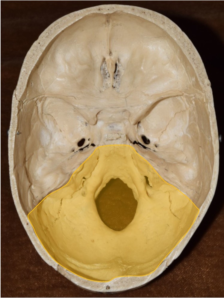
Cranial nerve foramina are integral exits from the confines of the skull. Despite their significance in cranial nerve pathologies, there has been no comprehensive anatomical review of these structures. Owing to the extensive nature of this topic we have divided our review into two parts; Part II, presented here, focuses on the foramina of the posterior cranial fossa and discusses each foramen's shape, orientation, size, surrounding structures, and structures that pass through it. Furthermore, by comparing foramen sizes against the cross-sectional areas of their contents, we determine the amount of free space available within each. We also review lesions that can obstruct each foramen and discuss the clinical consequences.

摘要

颅神经孔是颅骨范围内不可或缺的出口。尽管它们在颅神经病变中具有重要意义,但尚未对这些结构进行全面的解剖学综述。由于该主题范围广泛,我们将综述分为两部分;此处呈现的第二部分聚焦于后颅窝的孔,并讨论每个孔的形状、方向、大小、周围结构以及穿过它的结构。此外,通过将孔的大小与其内容物的横截面积进行比较,我们确定每个孔内可用的自由空间量。我们还综述了可能阻塞每个孔的病变并讨论其临床后果。

https://cdn.ncbi.nlm.nih.gov/pmc/blobs/94db/6005399/c19bb87e043c/cureus-0010-00000002500-i02.jpg
https://cdn.ncbi.nlm.nih.gov/pmc/blobs/94db/6005399/653f446fa26f/cureus-0010-00000002500-i01.jpg
https://cdn.ncbi.nlm.nih.gov/pmc/blobs/94db/6005399/c19bb87e043c/cureus-0010-00000002500-i02.jpg
https://cdn.ncbi.nlm.nih.gov/pmc/blobs/94db/6005399/653f446fa26f/cureus-0010-00000002500-i01.jpg
https://cdn.ncbi.nlm.nih.gov/pmc/blobs/94db/6005399/c19bb87e043c/cureus-0010-00000002500-i02.jpg

相似文献

1
Cranial Nerve Foramina: Part II - A Review of the Anatomy and Pathology of Cranial Nerve Foramina of the Posterior Cranial Fossa.颅神经孔:第二部分 - 后颅窝颅神经孔的解剖学与病理学综述
Cureus. 2018 Apr 18;10(4):e2500. doi: 10.7759/cureus.2500.
2
Cranial Nerve Foramina Part I: A Review of the Anatomy and Pathology of Cranial Nerve Foramina of the Anterior and Middle Fossa.颅神经孔 第一部分:前颅窝和中颅窝颅神经孔的解剖与病理学综述
Cureus. 2018 Feb 8;10(2):e2172. doi: 10.7759/cureus.2172.
3
Variant Bilateral Foramina of the Middle Cranial Fossa.颅中窝双侧变异孔
Cureus. 2022 Dec 27;14(12):e33014. doi: 10.7759/cureus.33014. eCollection 2022 Dec.
4
Endonasal access to lower cranial nerves: From foramina to upper parapharyngeal space.经鼻颅神经入路:从神经孔到咽旁上间隙。
Head Neck. 2021 Oct;43(10):3225-3233. doi: 10.1002/hed.26781. Epub 2021 Jun 24.
5
Visualization of Dark Side of Skull Base with Surgical Navigation and Endoscopic Assistance: Extended Petrous Rhomboid and Rhomboid with Maxillary Nerve-Mandibular Nerve Vidian Corridor.手术导航和内镜辅助下颅底黑暗面的可视化:扩展的岩枕部和岩枕部,以及上颌神经-下颌神经翼管。
World Neurosurg. 2019 Sep;129:e134-e145. doi: 10.1016/j.wneu.2019.05.062. Epub 2019 May 17.
6
Occipital foramina development involves localised regulation of mesenchyme proliferation and is independent of apoptosis.枕骨大孔的发育涉及间充质增殖的局部调节,且与细胞凋亡无关。
J Anat. 2015 Jun;226(6):560-74. doi: 10.1111/joa.12304.
7
Foramina and canals of skull base in Holstein cattle: a computed tomography study.荷斯坦奶牛颅底的孔和管:一项计算机断层扫描研究。
Folia Morphol (Warsz). 2025;84(1):216-230. doi: 10.5603/fm.100461. Epub 2024 Jun 10.
8
Jugular Foramen Syndrome颈静脉孔综合征
9
The variable relationship between the lower cranial nerves and jugular foramen tumors: implications for neural preservation.下颅神经与颈静脉孔区肿瘤之间的可变关系:对神经保留的意义。
Am J Otol. 1996 Jul;17(4):658-68.
10
Microsurgical anatomy for lateral approaches to the foramen magnum with special reference to transcondylar fossa (supracondylar transjugular tubercle) approach.枕骨大孔外侧入路的显微外科解剖学,特别参考髁后窝(髁上经颈静脉结节)入路。
Skull Base Surg. 1998;8(3):119-25. doi: 10.1055/s-2008-1058570.

引用本文的文献

1
Case report: A rare case of neurocytoma of the Vth cranial nerve.病例报告:一例罕见的第五颅神经神经细胞瘤。
Front Oncol. 2024 Sep 27;14:1438011. doi: 10.3389/fonc.2024.1438011. eCollection 2024.
2
Osteoclast activity sculpts craniofacial form to permit sensorineural patterning in the zebrafish skull.破骨细胞的活动塑造了颅面形态,以允许在斑马鱼颅骨中进行感觉神经模式形成。
Front Endocrinol (Lausanne). 2022 Nov 1;13:969481. doi: 10.3389/fendo.2022.969481. eCollection 2022.
3
Biomechanical analysis of skull trauma and opportunity in neuroradiology interpretation to explain the post-concussion syndrome: literature review and case studies presentation.

本文引用的文献

1
Morphological and Morphometric Analysis of Hypoglossal Canal in North Indian Dry Skulls and It's Significance in Cranial Base Surgeries.北印度干燥颅骨舌下神经管的形态学和形态测量分析及其在颅底手术中的意义。
J Clin Diagn Res. 2017 Mar;11(3):AC08-AC12. doi: 10.7860/JCDR/2017/24333.9365. Epub 2017 Mar 1.
2
Foramen magnum, occipital condyles and hypoglossal canals morphometry: anatomical study with clinical implications.枕骨大孔、枕髁及舌下神经管形态测量:具有临床意义的解剖学研究
Folia Morphol (Warsz). 2017;76(3):446-457. doi: 10.5603/FM.a2017.0002. Epub 2017 Feb 2.
3
Morphology, topography and clinical signi cance of the jugular foramen.
颅骨创伤的生物力学分析以及神经放射学解读中解释脑震荡后综合征的机会:文献综述与病例研究展示
Eur Radiol Exp. 2020 Dec 8;4(1):66. doi: 10.1186/s41747-020-00194-x.
4
Lower cranial nerve syndromes: a review.颅神经综合征:综述。
Neurosurg Rev. 2021 Jun;44(3):1345-1355. doi: 10.1007/s10143-020-01344-w. Epub 2020 Jul 8.
Folia Med Cracov. 2016;56(1):71-80.
4
Foramen magnum dural arteriovenous fistula presenting with epilepsy.以癫痫为表现的枕骨大孔区硬脑膜动静脉瘘
Interv Neuroradiol. 2015 Dec;21(6):724-7. doi: 10.1177/1591019915609783. Epub 2015 Oct 15.
5
Microsurgical Anatomy of the Labyrinthine Artery and Clinical Relevance.迷路动脉的显微外科解剖及临床意义
Turk Neurosurg. 2015;25(4):539-43. doi: 10.5137/1019-5149.JTN.9136-13.0.
6
Meningioma of foramen magnum causing drop attacks.枕骨大孔脑膜瘤导致猝倒发作。
Case Rep Neurol Med. 2015;2015:214563. doi: 10.1155/2015/214563. Epub 2015 Feb 22.
7
Labyrinthine artery aneurysm as an internal auditory canal mass.迷路动脉动脉瘤表现为内耳道肿块。
J Neurol Surg Rep. 2014 Aug;75(1):e38-41. doi: 10.1055/s-0033-1358796. Epub 2013 Dec 12.
8
A case of Vernet syndrome associated with internal jugular phlebectasia.1例伴颈内静脉扩张的韦尔内综合征。
PM R. 2014 Dec;6(12):1163-5. doi: 10.1016/j.pmrj.2014.05.025. Epub 2014 Jul 1.
9
Osteoma of the internal auditory canal mimicking vestibular schwannoma: case report and review of 17 recent cases.内听道骨瘤表现为前庭神经鞘瘤:病例报告及对 17 例近期病例的回顾。
Tohoku J Exp Med. 2014 Jan;232(1):63-8. doi: 10.1620/tjem.232.63.
10
Establishing normal diameter range of the cochlear and facial nerves with 3D-CISS at 3T.建立 3T 下 3D-CISS 耳蜗神经和面神经正常直径范围。
Magn Reson Med Sci. 2013 Dec 25;12(4):241-7. doi: 10.2463/mrms.2013-0004. Epub 2013 Oct 29.