Bioartificial organs, Biomaterials Science and Technology, MIRA Institute of Biomedical Technology and Technical Medicine, University of Twente, Enschede, The Netherlands.
Developmental BioEngineering, MIRA Institute of Biomedical Technology and Technical Medicine, University of Twente, Enschede, The Netherlands.
J Mater Sci Mater Med. 2018 Jun 25;29(7):91. doi: 10.1007/s10856-018-6102-0.
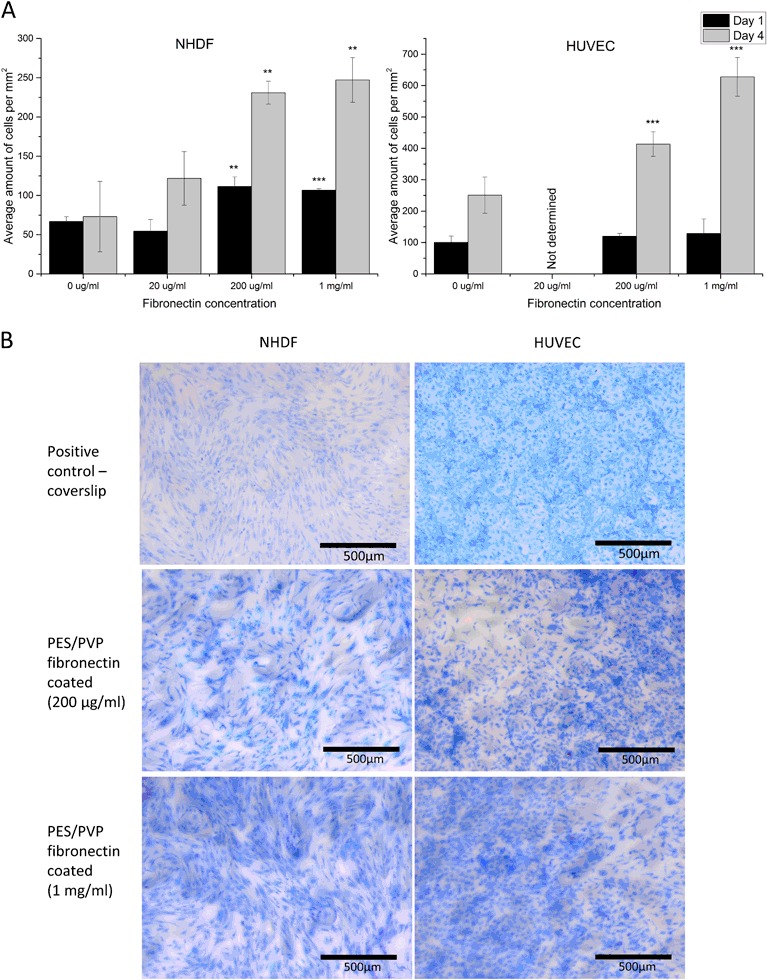
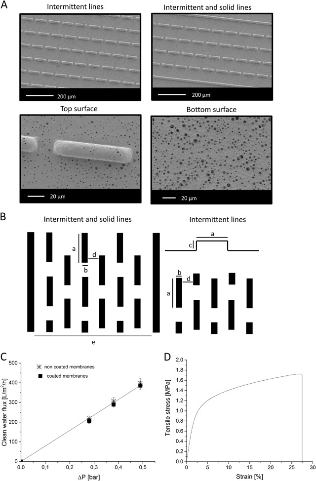
The development of immune protective islet encapsulation devices could allow for islet transplantation in the absence of immunosuppression. However, the immune protective membrane / barrier introduced there could also impose limitations in transport of oxygen and nutrients to the encapsulated cells resulting to limited islet viability. In the last years, it is well understood that achieving prevascularization of the device in vitro could facilitate its connection to the host vasculature after implantation, and therefore could provide sufficient blood supply and oxygenation to the encapsulated islets. However, the microvascular networks created in vitro need to mimic well the highly organized vasculature of the native tissue. In earlier study, we developed a functional macroencapsulation device consisting of two polyethersulfone/polyvinylpyrrolidone (PES/PVP) membranes, where a bottom microwell membrane provides good separation of encapsulated islets and the top flat membrane acts as a lid. In this work, we investigate the possibility of creating early microvascular networks on the lid of this device by combining novel membrane microfabrication with co-culture of human umbilical vein endothelial cell (HUVEC) and fibroblasts. We create thin porous microstructured PES/PVP membranes with solid and intermittent line-patterns and investigate the effect of cell alignment and cell interconnectivity as a first step towards the development of a stable prevascularized layer in vitro. Our results show that, in contrast to non-patterned membranes where HUVECs form unorganized HUVEC branch-like structures, for the micropatterned membranes, we can achieve cell alignment and the co-culture of HUVECs on a monolayer of fibroblasts attached on the membranes with intermittent line-pattern allows for the creation of HUVEC branch-like structures over the membrane surface. This important step towards creating early microvascular networks was achieved without the addition of hydrogels, often used in angiogenesis assays, as gels could block the pores of the membrane and limit the transport properties of the islet encapsulation device.
免疫保护胰岛包封装置的发展可以实现无免疫抑制的胰岛移植。然而,引入的免疫保护膜/屏障也可能对包裹细胞的氧气和营养物质的传输造成限制,从而导致包裹胰岛的活力有限。在过去几年中,人们已经充分认识到,在体外实现装置的预血管化可以促进其在植入后的与宿主血管的连接,从而为包裹的胰岛提供足够的血液供应和氧合。然而,体外创建的微血管网络需要很好地模拟天然组织中高度组织化的脉管系统。在早期的研究中,我们开发了一种由两层聚醚砜/聚乙烯吡咯烷酮(PES/PVP)膜组成的功能性宏观包封装置,其中底层微孔膜提供了对包裹胰岛的良好分离,而顶层平板膜作为盖子。在这项工作中,我们通过将新型膜微加工与人类脐静脉内皮细胞(HUVEC)和成纤维细胞的共培养相结合,研究了在该装置的盖子上创建早期微血管网络的可能性。我们创建了具有实心和间断线图案的薄多孔 PES/PVP 膜,并研究了细胞取向和细胞连通性的影响,作为在体外开发稳定预血管化层的第一步。我们的结果表明,与 HUVEC 形成无组织的 HUVEC 分支状结构的非图案化膜相反,对于微图案化膜,我们可以实现细胞取向,并且 HUVEC 可以在附着于膜上的成纤维细胞单层上共培养,并且在膜表面上允许形成 HUVEC 分支状结构的间断线图案允许在膜表面上形成 HUVEC 分支状结构。在没有添加经常用于血管生成测定的水凝胶的情况下,实现了这一朝着创建早期微血管网络的重要步骤,因为凝胶可能会堵塞膜的孔并限制胰岛包封装置的传输特性。