Section for Translational Psychobiology in Child and Adolescent Psychiatry, Department of Child and Adolescent Psychiatry, Centre for Psychosocial Medicine, University of Heidelberg, Heidelberg, Germany.
University Hospital of Child and Adolescent Psychiatry and Psychotherapy, University of Bern, Bern 60, Switzerland.
Soc Cogn Affect Neurosci. 2018 Sep 4;13(7):741-753. doi: 10.1093/scan/nsy046.
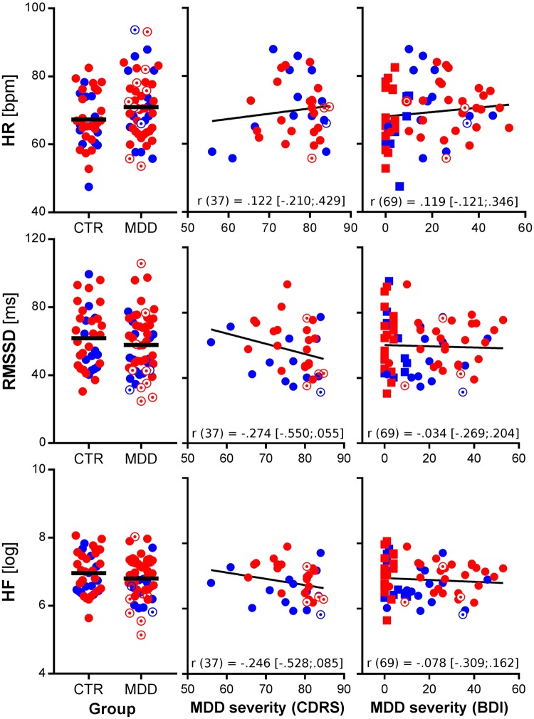
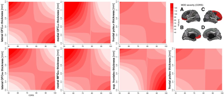
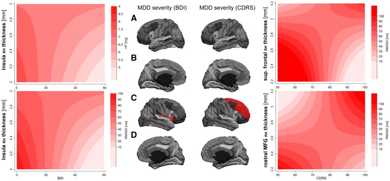
Major depressive disorder (MDD) has been associated with abnormalities in cortical thickness and autonomic function. Adolescence is a time notable for brain development and MDD onset. In healthy adolescents, greater resting state vagal activity (RVA) is associated with lower cortical thickness. The relationship between brain structural thickness and RVA in adolescents with MDD has not previously been studied. This secondary analysis drew on a sample of 37 non-depressed controls and 53 adolescents with MDD. Resting state heart rate and two indices of RVA (HF-HRV and RMSSD) were recorded during a neuroimaging session. Cortical thickness within fronto-limbic regions of interest was measured using Freesurfer analysis of T1-weighted high-resolution structural images. Self-reports of depression severity showed a significant interaction with cortical thickness of the right insula in predicting RMSSD [t = 2.22, P=0.030, β = 5.44; model fit of the interaction term as indicated by the 'Bayes Factor' (BF): 7.58] and HF-HRV (t = 2.09, P=0.041, β = 4.72; BF: 7.94). Clinician ratings of depression severity showed further interactions. Findings underscore the important relationships between RVA and cortical development, suggesting two possible explanations: (i) in adolescent MDD, greater fronto-limbic thickness is compensatory for deficits in autonomic regulation or (ii) increased autonomic arousal results in delayed fronto-limbic maturation. Longitudinal research is necessary to further clarify the nature of the relationship between autonomic functioning and cortical development.
重度抑郁症(MDD)与皮质厚度和自主功能异常有关。青春期是大脑发育和 MDD 发病的重要时期。在健康的青少年中,静息状态下迷走神经活动(RVA)越高,皮质厚度越低。MDD 青少年的大脑结构厚度与 RVA 之间的关系以前尚未研究过。这项二次分析基于 37 名非抑郁对照组和 53 名 MDD 青少年的样本。在神经影像学检查期间记录了静息状态下的心率和 RVA 的两个指标(HF-HRV 和 RMSSD)。使用 Freesurfer 对 T1 加权高分辨率结构图像进行分析,测量额-边缘区域感兴趣区内的皮质厚度。抑郁严重程度的自我报告与右侧岛叶皮质厚度在预测 RMSSD 方面存在显著的交互作用[t=2.22,P=0.030,β=5.44;交互项的“贝叶斯因子”(BF)表示模型拟合度:7.58]和 HF-HRV(t=2.09,P=0.041,β=4.72;BF:7.94)。临床医生对抑郁严重程度的评估显示出进一步的相互作用。这些发现强调了 RVA 和皮质发育之间的重要关系,表明了两种可能的解释:(i)在青少年 MDD 中,额-边缘厚度的增加是对自主调节缺陷的补偿,或(ii)自主唤醒增加导致额-边缘成熟延迟。需要进行纵向研究以进一步阐明自主功能与皮质发育之间关系的本质。