• 文献检索
  • 文档翻译
  • 深度研究
  • 学术资讯
  • Suppr Zotero 插件Zotero 插件
  • 邀请有礼
  • 套餐&价格
  • 历史记录
应用&插件
Suppr Zotero 插件Zotero 插件浏览器插件Mac 客户端Windows 客户端微信小程序
定价
高级版会员购买积分包购买API积分包
服务
文献检索文档翻译深度研究API 文档MCP 服务
关于我们
关于 Suppr公司介绍联系我们用户协议隐私条款
关注我们

Suppr 超能文献

核心技术专利:CN118964589B侵权必究
粤ICP备2023148730 号-1Suppr @ 2026

文献检索

告别复杂PubMed语法,用中文像聊天一样搜索,搜遍4000万医学文献。AI智能推荐,让科研检索更轻松。

立即免费搜索

文件翻译

保留排版,准确专业,支持PDF/Word/PPT等文件格式,支持 12+语言互译。

免费翻译文档

深度研究

AI帮你快速写综述,25分钟生成高质量综述,智能提取关键信息,辅助科研写作。

立即免费体验

评估 T2-W MR 成像和弥散加权成像在宫颈癌保妇治疗后早期局部反应评估中的应用:一项多中心研究。

Evaluation of T2-W MR imaging and diffusion-weighted imaging for the early post-treatment local response assessment of patients treated conservatively for cervical cancer: a multicentre study.

机构信息

Department of Radiology and Nuclear Medicine, Erasmus University Medical Center, Rotterdam, The Netherlands.

Department of Radiology, Erasmus Medical Center Rotterdam, P.O Box 2040, 3015, CE, Rotterdam, The Netherlands.

出版信息

Eur Radiol. 2019 Jan;29(1):309-318. doi: 10.1007/s00330-018-5510-3. Epub 2018 Jun 25.

DOI:10.1007/s00330-018-5510-3
PMID:29943182
原文链接:https://pmc.ncbi.nlm.nih.gov/articles/PMC6291430/
Abstract

OBJECTIVES

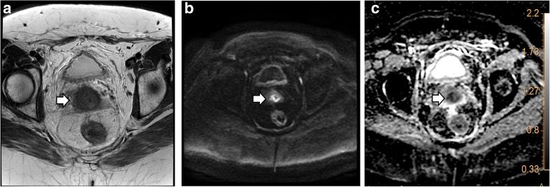
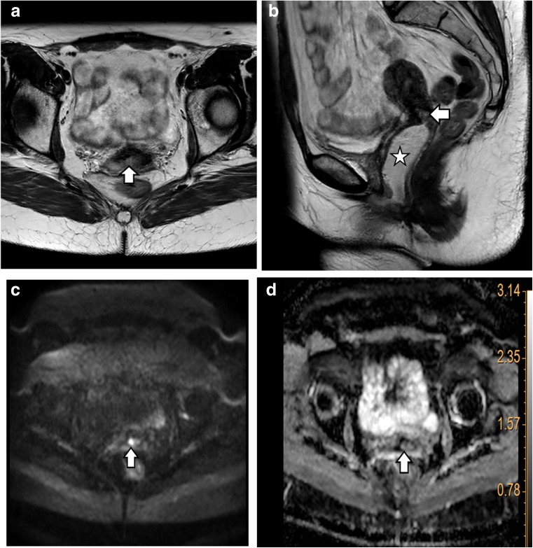
To compare MR imaging with or without DWI and clinical response evaluation (CRE) in the local control evaluation of cervical carcinoma after radiotherapy.

METHODS

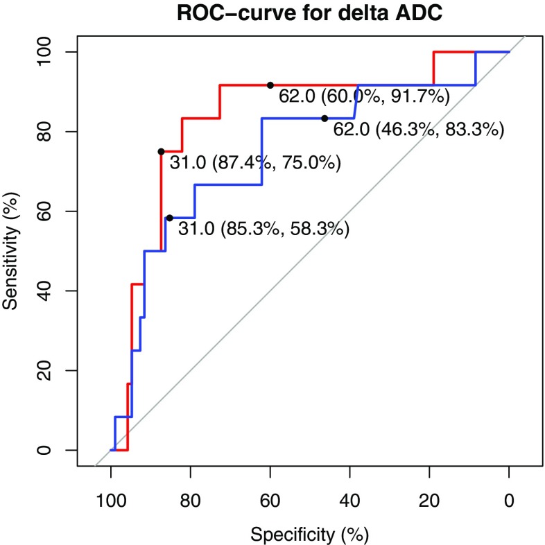
In a multicentre university setting, we prospectively included 107 patients with primary cervical cancer treated with radiotherapy. Sensitivity and specificity for CRE and MR imaging (with pre-therapy MR imaging as reference) (2 readers) were evaluated using cautious and strict criteria for identifying residual tumour. Nested logistic regression models were constructed for CRE, subsequently adding MR imaging with and without DWI as independent variables, as well as the pre- to post-treatment change in apparent diffusion coefficient (delta ADC).

RESULTS

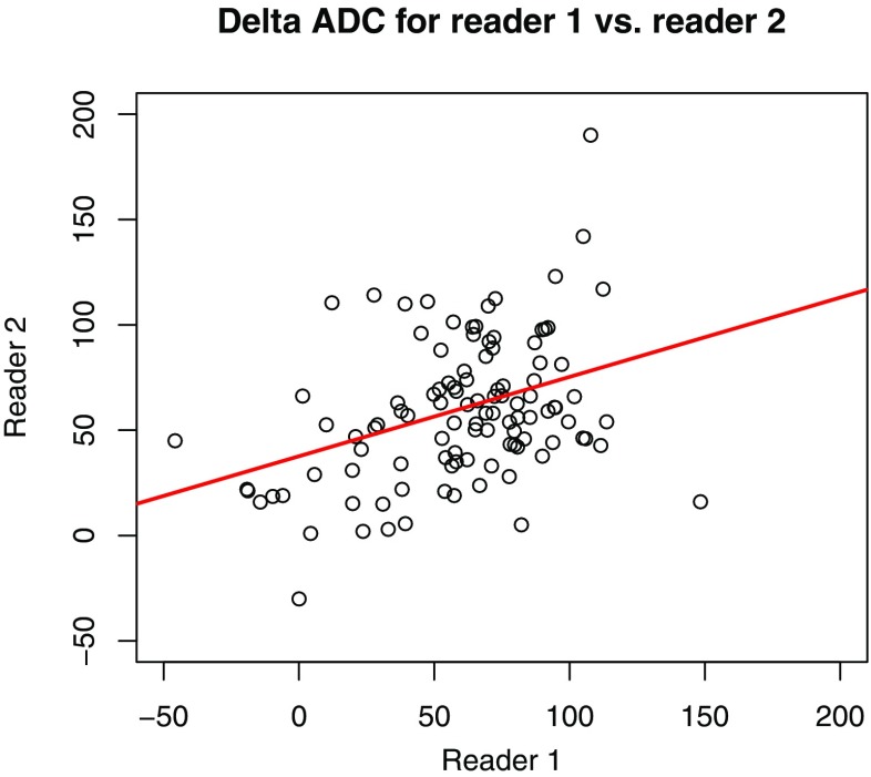
Using cautious criteria, CRE and MR imaging with DWI (reader 1/reader 2) have comparable high specificity (83% and 89%/95%, respectively), whereas MR imaging without DWI showed significantly lower specificity (63%/53%) than CRE. Using strict criteria, CRE and MR imaging with DWI both showed very high specificity (99% and 92%/95%, respectively), whereas MR imaging without DWI showed significantly lower specificity (89%/77%) than CRE. All sensitivities were not significantly different. Addition of MR imaging with DWI to CRE has statistically significant incremental value in identifying residual tumour (reader 1: estimate, 1.06; p = 0.001) (reader 2: estimate, 0.62; p = 0.02). Adding the delta ADC did not have significant incremental value in detecting residual tumour.

CONCLUSIONS

DWI significantly increases the specificity of MR imaging in the detection of local residual tumour. Furthermore, MR imaging with DWI has significant incremental diagnostic value over CRE, whereas adding the delta ADC has no incremental diagnostic value.

KEY POINTS

• If MR imaging is used for response evaluation, DWI should be incorporated • MR imaging with DWI has diagnostic value comparable/complementary to clinical response evaluation • Inter-reader agreement is moderate to fair for two experienced radiologist readers • Quantitative measurements of ADC early post-therapy have limited diagnostic value.

摘要

目的

比较磁共振成像(MR 成像)加或不加弥散加权成像(DWI)与临床反应评估(CRE)在宫颈癌放疗后局部控制评估中的作用。

方法

在多中心大学环境中,我们前瞻性地纳入了 107 例接受放疗的原发性宫颈癌患者。使用谨慎和严格的标准来识别残留肿瘤,评估 CRE 和 MR 成像(以治疗前的 MR 成像作为参考)(2 位读者)的敏感性和特异性。构建嵌套逻辑回归模型,将 CRE 作为因变量,MR 成像加或不加 DWI 以及表观扩散系数(ADC)的治疗前后变化(delta ADC)作为独立变量。

结果

使用谨慎标准,CRE 和 MR 成像加 DWI(读者 1/读者 2)的特异性具有可比性(分别为 83%和 89%/95%),而不加 DWI 的 MR 成像特异性显著较低(分别为 63%/53%)。使用严格标准,CRE 和 MR 成像加 DWI 均显示出非常高的特异性(分别为 99%和 92%/95%),而不加 DWI 的 MR 成像特异性显著较低(分别为 89%/77%)。所有的敏感性均无显著差异。MR 成像加 DWI 与 CRE 联合应用具有统计学意义的识别残留肿瘤的增量价值(读者 1:估计值,1.06;p = 0.001)(读者 2:估计值,0.62;p = 0.02)。添加 delta ADC 对检测残留肿瘤没有显著的增量价值。

结论

DWI 显著提高了 MR 成像检测局部残留肿瘤的特异性。此外,MR 成像加 DWI 比 CRE 具有显著的增量诊断价值,而添加 delta ADC 则没有增量诊断价值。

关键点

  • 如果使用 MR 成像进行反应评估,则应纳入 DWI。

  • MR 成像加 DWI 具有与临床反应评估相当/互补的诊断价值。

  • 两位有经验的放射科医生读者的读者间一致性为中等至良好。

  • 治疗后早期 ADC 的定量测量值具有有限的诊断价值。

https://cdn.ncbi.nlm.nih.gov/pmc/blobs/b74f/6291430/bd811893289e/330_2018_5510_Fig4_HTML.jpg
https://cdn.ncbi.nlm.nih.gov/pmc/blobs/b74f/6291430/d80494785a4d/330_2018_5510_Fig1_HTML.jpg
https://cdn.ncbi.nlm.nih.gov/pmc/blobs/b74f/6291430/f050ca3bc6c1/330_2018_5510_Fig2_HTML.jpg
https://cdn.ncbi.nlm.nih.gov/pmc/blobs/b74f/6291430/83d54c21ec69/330_2018_5510_Fig3_HTML.jpg
https://cdn.ncbi.nlm.nih.gov/pmc/blobs/b74f/6291430/bd811893289e/330_2018_5510_Fig4_HTML.jpg

相似文献

1
Evaluation of T2-W MR imaging and diffusion-weighted imaging for the early post-treatment local response assessment of patients treated conservatively for cervical cancer: a multicentre study.评估 T2-W MR 成像和弥散加权成像在宫颈癌保妇治疗后早期局部反应评估中的应用:一项多中心研究。
Eur Radiol. 2019 Jan;29(1):309-318. doi: 10.1007/s00330-018-5510-3. Epub 2018 Jun 25.
2
Value of diffusion-weighted MRI in diagnosis of uterine cervical cancer: a prospective study evaluating the benefits of DWI compared to conventional MR sequences in a 3T environment.扩散加权磁共振成像在子宫颈癌诊断中的价值:一项前瞻性研究,评估在3T环境下弥散加权成像(DWI)与传统磁共振序列相比的优势。
Acta Radiol. 2016 Jul;57(7):869-77. doi: 10.1177/0284185115602146. Epub 2015 Sep 1.
3
Prediction of disease progression following concurrent chemoradiotherapy for uterine cervical cancer: value of post-treatment diffusion-weighted imaging.同步放化疗后子宫颈癌疾病进展的预测:治疗后扩散加权成像的价值
Eur Radiol. 2016 Sep;26(9):3272-9. doi: 10.1007/s00330-015-4156-7. Epub 2015 Dec 18.
4
[Value of MR diffusion-weighted imaging in diagnosis and outcome prediction for uterine cervical cancer].[磁共振扩散加权成像在子宫颈癌诊断及预后预测中的价值]
Zhonghua Zhong Liu Za Zhi. 2014 Feb;36(2):115-9.
5
Endometrial cancer with cervical stromal invasion: diagnostic accuracy of diffusion-weighted and dynamic contrast enhanced MR imaging at 3T.伴有宫颈间质浸润的子宫内膜癌:3T磁共振成像中扩散加权成像与动态对比增强成像的诊断准确性
Eur Radiol. 2017 May;27(5):1867-1876. doi: 10.1007/s00330-016-4583-0. Epub 2016 Sep 9.
6
Comparison of optimised endovaginal vs external array coil T2-weighted and diffusion-weighted imaging techniques for detecting suspected early stage (IA/IB1) uterine cervical cancer.优化的经阴道与体外阵列线圈T2加权和扩散加权成像技术在检测疑似早期(IA/IB1期)子宫颈癌中的比较。
Eur Radiol. 2016 Apr;26(4):941-50. doi: 10.1007/s00330-015-3899-5. Epub 2015 Jul 11.
7
Accuracy of diffusion-weighted echo-planar MR imaging and ADC mapping in the evaluation of residual cervical carcinoma after radiation therapy.弥散加权回波平面磁共振成像及 ADC 图在评估宫颈癌放疗后残留灶中的准确性。
Gynecol Oncol. 2011 Oct;123(1):110-5. doi: 10.1016/j.ygyno.2011.06.009. Epub 2011 Jul 20.
8
Evaluation of diffusion-weighted imaging (DWI) and MR spectroscopy (MRS) as early response biomarkers in cervical cancer patients.评估扩散加权成像(DWI)和磁共振波谱(MRS)作为宫颈癌患者早期反应生物标志物的情况。
Radiol Med. 2016 Nov;121(11):838-846. doi: 10.1007/s11547-016-0665-y. Epub 2016 Jul 2.
9
Diffusion-weighted magnetic resonance imaging in the early detection of response to chemoradiation in cervical cancer.扩散加权磁共振成像在宫颈癌放化疗反应早期检测中的应用
Gynecol Oncol. 2008 Nov;111(2):213-20. doi: 10.1016/j.ygyno.2008.07.048. Epub 2008 Sep 6.
10
The Value of Diffusion-Weighted Imaging in Combination With Conventional Magnetic Resonance Imaging for Improving Tumor Detection for Early Cervical Carcinoma Treated With Fertility-Sparing Surgery.扩散加权成像联合传统磁共振成像在提高保留生育功能手术治疗早期宫颈癌肿瘤检测中的价值
Int J Gynecol Cancer. 2017 Oct;27(8):1761-1768. doi: 10.1097/IGC.0000000000001113.

引用本文的文献

1
Multi-parametric MRI-based radiomics nomogram for predicting lymphovascular space invasion in early-stage cervical adenocarcinoma.基于多参数磁共振成像的影像组学列线图预测早期宫颈腺癌的淋巴血管间隙浸润
Front Oncol. 2025 Aug 21;15:1612691. doi: 10.3389/fonc.2025.1612691. eCollection 2025.
2
Prediction of Patient Outcomes in Locally Advanced Cervical Carcinoma Following Chemoradiotherapy-Comparative Effectiveness of Magnetic Resonance Imaging and 2-Deoxy-2-[F]fluoro-D-glucose Imaging.局部晚期宫颈癌放化疗后患者预后的预测——磁共振成像与2-脱氧-2-[F]氟代-D-葡萄糖成像的比较效能
Cancers (Basel). 2024 Jan 23;16(3):476. doi: 10.3390/cancers16030476.
3

本文引用的文献

1
Value of Dynamic Contrast-enhanced and Diffusion-weighted MR Imaging in the Detection of Pathologic Complete Response in Cervical Cancer after Neoadjuvant Therapy: A Retrospective Observational Study.动态对比增强磁共振成像和弥散加权成像在新辅助治疗后宫颈癌病理完全缓解检测中的价值:一项回顾性观察性研究。
Radiology. 2017 Aug;284(2):432-442. doi: 10.1148/radiol.2017161299. Epub 2017 Mar 16.
2
Follow-up for cervical cancer: a Program in Evidence-Based Care systematic review and clinical practice guideline update.子宫颈癌的随访:循证护理计划的系统评价与临床实践指南更新
Curr Oncol. 2016 Apr;23(2):109-18. doi: 10.3747/co.23.2742. Epub 2016 Apr 13.
3
Staging of Cervical Cancer: A Practical Approach Using MRI and FDG PET.
宫颈癌分期:MRI 和 FDG PET 的实用方法。
AJR Am J Roentgenol. 2023 Nov;221(5):633-648. doi: 10.2214/AJR.23.29003. Epub 2023 Jun 7.
4
Diagnostic Accuracy of Magnetic Resonance Imaging in the Pre-Operative Staging of Cervical Cancer Patients Who Underwent Neoadjuvant Treatment: A Clinical-Surgical-Pathologic Comparison.接受新辅助治疗的宫颈癌患者术前分期中磁共振成像的诊断准确性:临床-手术-病理比较
Cancers (Basel). 2023 Mar 30;15(7):2061. doi: 10.3390/cancers15072061.
5
A feasibility study of reduced full-of-view synthetic high-b-value diffusion-weighted imaging in uterine tumors.子宫肿瘤中减少全视野合成高b值扩散加权成像的可行性研究。
Insights Imaging. 2023 Jan 16;14(1):12. doi: 10.1186/s13244-022-01350-0.
6
Post treatment imaging in patients with local advanced cervical carcinoma.局部晚期宫颈癌患者的治疗后影像学检查
Front Oncol. 2022 Oct 27;12:1003930. doi: 10.3389/fonc.2022.1003930. eCollection 2022.
7
Magnetic resonance imaging in cervical cancer interventional radiotherapy (brachytherapy): a pictorial essay focused on radiologist management.宫颈癌介入放疗(近距离放疗)中的磁共振成像:一篇聚焦于放射科医生管理的图文并茂的文章
J Contemp Brachytherapy. 2022 Jun;14(3):287-298. doi: 10.5114/jcb.2022.117727. Epub 2022 Jun 30.
8
Early clinical outcomes of hybrid brachytherapy for locally advanced cervical cancer: making adverse situations in a favorable scenario.局部晚期宫颈癌近距离混合治疗的早期临床结果:在有利情况下应对不利情况。
J Contemp Brachytherapy. 2022 Aug;14(4):321-331. doi: 10.5114/jcb.2022.118831. Epub 2022 Aug 17.
9
An MRI-based radiomics signature and clinical characteristics for survival prediction in early-stage cervical cancer.基于 MRI 的放射组学特征和临床特征预测早期宫颈癌患者的生存情况。
Br J Radiol. 2022 Jan 1;95(1129):20210838. doi: 10.1259/bjr.20210838. Epub 2021 Nov 29.
10
Quantitative imaging of uterine cancers with diffusion-weighted MRI and 18-fluorodeoxyglucose PET/CT.用弥散加权 MRI 和 18-氟脱氧葡萄糖 PET/CT 对子宫癌进行定量成像。
Abdom Radiol (NY). 2022 Sep;47(9):3174-3188. doi: 10.1007/s00261-021-03218-1. Epub 2021 Jul 23.
Prediction of disease progression following concurrent chemoradiotherapy for uterine cervical cancer: value of post-treatment diffusion-weighted imaging.
同步放化疗后子宫颈癌疾病进展的预测:治疗后扩散加权成像的价值
Eur Radiol. 2016 Sep;26(9):3272-9. doi: 10.1007/s00330-015-4156-7. Epub 2015 Dec 18.
4
Curative pelvic exenteration for recurrent cervical carcinoma in the era of concurrent chemotherapy and radiation therapy. A systematic review.同步放化疗时代复发性宫颈癌的盆腔根治性脏器切除术:一项系统评价
Eur J Surg Oncol. 2015 Aug;41(8):975-85. doi: 10.1016/j.ejso.2015.03.235. Epub 2015 Apr 14.
5
Role of the Apparent Diffusion Coefficient in the Prediction of Response to Neoadjuvant Chemotherapy in Patients With Locally Advanced Breast Cancer.表观扩散系数在局部晚期乳腺癌患者新辅助化疗反应预测中的作用
Clin Breast Cancer. 2015 Oct;15(5):370-80. doi: 10.1016/j.clbc.2015.02.002. Epub 2015 Feb 21.
6
Monitoring treatment response in patients undergoing chemoradiotherapy for locally advanced uterine cervical cancer by additional diffusion-weighted imaging: A systematic review.通过额外的扩散加权成像监测局部晚期子宫颈癌患者放化疗的治疗反应:一项系统评价。
J Magn Reson Imaging. 2015 Sep;42(3):572-94. doi: 10.1002/jmri.24784. Epub 2014 Oct 24.
7
Diffusion-weighted MR imaging of upper abdominal organs: field strength and intervendor variability of apparent diffusion coefficients.腹部器官的弥散加权磁共振成像:表观弥散系数的场强和不同供应商间的可变性。
Radiology. 2014 Feb;270(2):454-63. doi: 10.1148/radiol.13130819. Epub 2013 Nov 5.
8
Evaluation and impact of residual disease in locally advanced cervical cancer after concurrent chemoradiation therapy: results of a multicenter study.局部晚期宫颈癌同期放化疗后残留病灶的评估及其影响:多中心研究结果。
Eur J Surg Oncol. 2013 Dec;39(12):1428-34. doi: 10.1016/j.ejso.2013.10.006. Epub 2013 Oct 23.
9
MRI-guided adaptive radiotherapy in locally advanced cervical cancer from a Nordic perspective.MRI 引导自适应放疗在局部晚期宫颈癌治疗中的北欧视角。
Acta Oncol. 2013 Oct;52(7):1510-9. doi: 10.3109/0284186X.2013.818253. Epub 2013 Aug 21.
10
Clinical examination versus magnetic resonance imaging in the pretreatment staging of cervical carcinoma: systematic review and meta-analysis.临床检查与磁共振成像在宫颈癌术前分期中的比较:系统评价和荟萃分析。
Eur Radiol. 2013 Jul;23(7):2005-18. doi: 10.1007/s00330-013-2783-4. Epub 2013 Mar 1.