Bashiri Dezfouli Ali, Pourfathollah Ali Akbar, Salar-Amoli Jamileh, Khosravi Mohammad, Nikogoftar-Zarif Mahin, Yazdi Mina, Ali-Esfahani Tahereh
Department of Basic Sciences, Faculty of Veterinary Medicine, University of Tehran, Tehran, Iran.
Department of Immunology, Faculty of Medical Science, Tarbiat Modares University, Tehran, Iran.
Med J Islam Repub Iran. 2017 Dec 17;31:98. doi: 10.14196/mjiri.31.98. eCollection 2017.
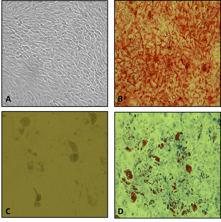
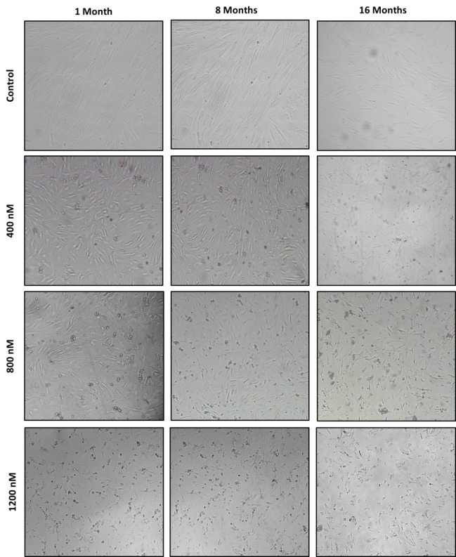
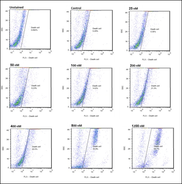
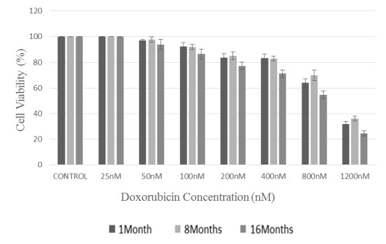
Doxorubicin, by aggregating in bone marrow, causes genotoxic effects, and thus reduces the repair ability of cells. The present study was conducted as an in vitro evaluation of age effects on the cytotoxicity induced by doxorubicin in mesenchymal stem cells (MSCs). The MSCs of female BALB/c mice aged 1, 8, and 16 months were separated, characterized, and subsequently evaluated in cellular growth media. After 24 hours, exposure of the MSCs of the 3 groups of mice to doxorubicin (25, 50, 100, 200, 400, 800, 1200 nM) and cytotoxicity were assessed, and the sublethal dose was determined using flow cytometry technique and lactate dehydrogenase (LDH) release assay. The IC50 values determined by flow cytometry for the separated MSCs of 1 young, 8 middle- aged, and 16 old mice were and respectively. Interestingly, the results of these 2 methods in determining cytotoxicity were in agreement, and a concentration of approximately 25 nM was considered to be the shared sublethal dose for different ages. The results indicated that MSCs of middle-aged mice were more resistant to the toxic effects of the drug. Besides, MSCs separated from the old mice were the most sensitive to chemotherapy and its side effects such as disruptions of cell proliferation and viability. These disruptions can be ascribed to the alteration of function and physiological processes with age. Determining proper concentration of doxorubicin drug to destruct cancerous cells based on age and individual sensitivity can minimize the amount of toxicity.
阿霉素通过在骨髓中聚集产生遗传毒性作用,从而降低细胞的修复能力。本研究旨在对阿霉素诱导间充质干细胞(MSCs)产生的细胞毒性的年龄效应进行体外评估。分离出1月龄、8月龄和16月龄雌性BALB/c小鼠的MSCs,进行特征鉴定,随后在细胞生长培养基中进行评估。24小时后,评估3组小鼠的MSCs暴露于阿霉素(25、50、100、200、400、800、1200 nM)后的细胞毒性,并使用流式细胞术和乳酸脱氢酶(LDH)释放测定法确定亚致死剂量。通过流式细胞术测定的1只年轻小鼠、8只中年小鼠和16只老年小鼠分离出的MSCs的IC50值分别为 。有趣的是,这两种确定细胞毒性方法的结果一致,约25 nM的浓度被认为是不同年龄的共同亚致死剂量。结果表明,中年小鼠的MSCs对该药物的毒性作用更具抗性。此外,从老年小鼠分离出的MSCs对化疗及其副作用(如细胞增殖和活力的破坏)最为敏感。这些破坏可归因于随着年龄增长功能和生理过程的改变。根据年龄和个体敏感性确定破坏癌细胞的阿霉素药物的合适浓度可以将毒性降至最低。