Division of Cardiovascular Medicine, Carver College of Medicine University of Iowa, Iowa City, IA
Division of Cardiovascular Medicine, Carver College of Medicine University of Iowa, Iowa City, IA.
J Am Heart Assoc. 2018 Jun 30;7(13):e006908. doi: 10.1161/JAHA.117.006908.
The epithelial growth factor receptor family of tyrosine kinases modulates embryonic formation of semilunar valves. We hypothesized that mice heterozygous for a dominant loss-of-function mutation in epithelial growth factor receptor, which are mice, would develop anomalous aortic valves, valve dysfunction, and valvular cardiomyopathy.
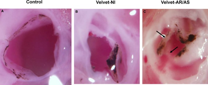
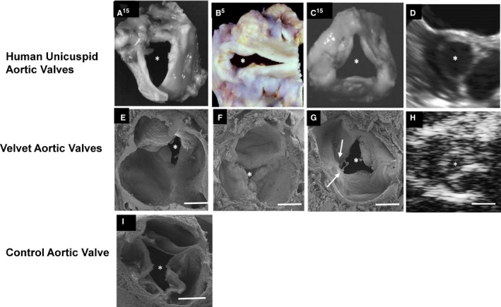
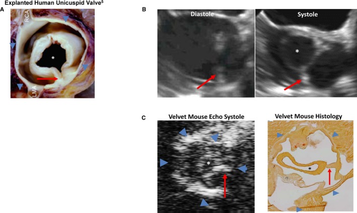
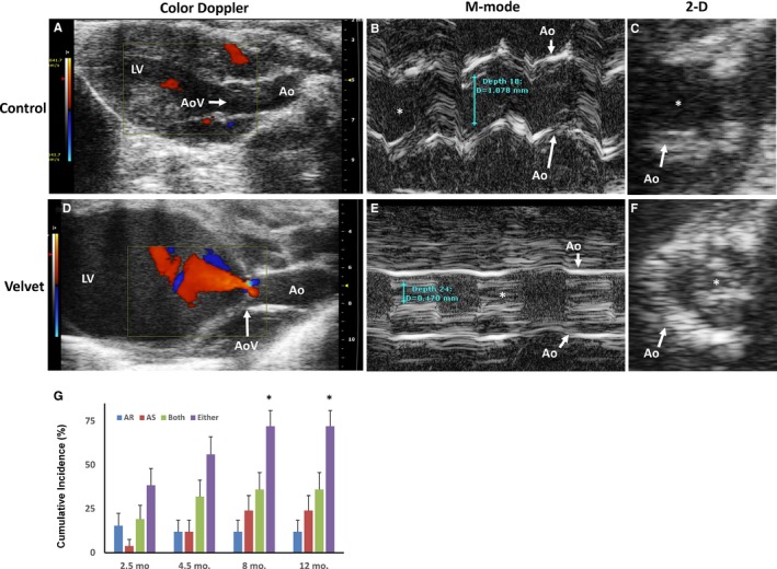
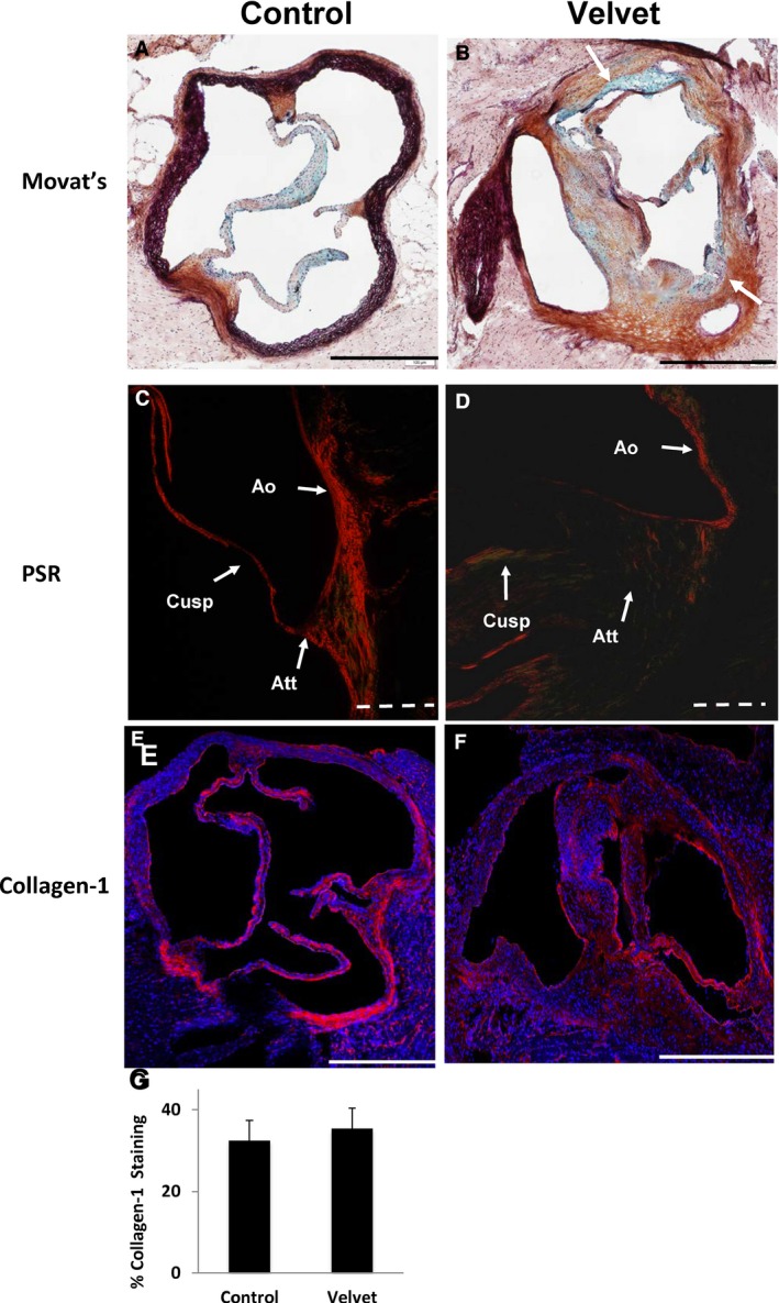
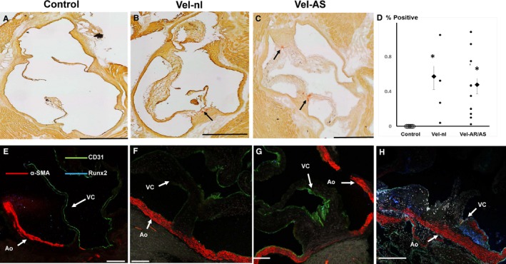
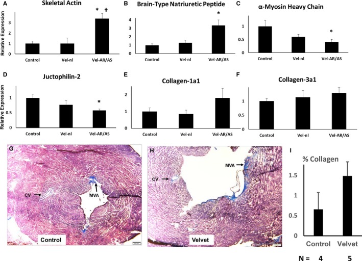
Aortic valves from mice and control mice were examined by light microscopy at 2.5 to 4 months of age. Additional and control mice underwent echocardiography at 2.5, 4.5, 8, and 12 months of age, followed by histologic examination. In young mice, microscopy revealed anatomic anomalies in 79% of aortic valves, which resembled human unicuspid aortic valves. Anomalies were not observed in control mice. At 12 months of age, histologic architecture was grossly distorted in aortic valves. Echocardiography detected moderate or severe aortic regurgitation, or aortic stenosis was present in 38% of mice at 2.5 months of age (N=24) and in 74% by 8 months of age. Left ventricular enlargement, hypertrophy, and reversion to a fetal myocardial gene expression program occurred in mice with aortic valve dysfunction, but not in mice with near-normal aortic valve function. Myocardial fibrosis was minimal or absent in all groups.
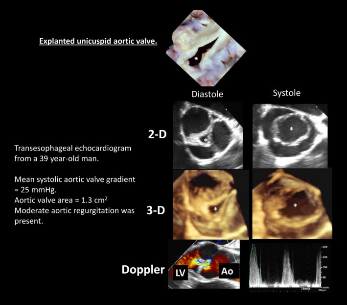
A new mouse model uniquely recapitulates salient functional, structural, and histologic features of human unicuspid aortic valve disease, which are phenotypically distinct from other forms of congenital aortic valve disease. The new model may be useful for elucidating mechanisms by which congenitally anomalous aortic valves become critically dysfunctional.
表皮生长因子受体家族的酪氨酸激酶调节半月瓣的胚胎形成。我们假设,表皮生长因子受体杂合显性失活突变的小鼠,即 小鼠,会发展出异常的主动脉瓣、瓣功能障碍和瓣性心肌病。
在 2.5 至 4 个月大时,通过光镜检查 小鼠和对照小鼠的主动脉瓣。另外一些 和对照小鼠在 2.5、4.5、8 和 12 个月大时进行超声心动图检查,然后进行组织学检查。在幼鼠中,显微镜检查发现 79%的 主动脉瓣存在解剖异常,类似于人类的单瓣主动脉瓣。在对照小鼠中未观察到异常。在 12 个月大时, 主动脉瓣的组织学结构严重扭曲。超声心动图在 2.5 个月大时(N=24)检测到 38%的 小鼠存在中度或重度主动脉瓣反流,或主动脉瓣狭窄,在 8 个月大时这一比例增加到 74%。在主动脉瓣功能障碍的 小鼠中出现左心室扩张、肥大和向胎儿心肌基因表达程序的逆转,但在主动脉瓣功能接近正常的 小鼠中没有出现这种情况。在所有组中,心肌纤维化都很少或不存在。
一种新的小鼠模型独特地再现了人类单瓣主动脉瓣疾病的显著功能、结构和组织学特征,其表型与其他形式的先天性主动脉瓣疾病明显不同。该新模型可能有助于阐明先天性异常主动脉瓣如何变得严重功能失调的机制。