Ueda Yoshitomo, Bando Yoshio, Misumi Sachiyo, Ogawa Shino, Ishida Akimasa, Jung Cha-Gyun, Shimizu Takeshi, Hida Hideki
Department of Neurophysiology and Brain Science, Nagoya City University Graduate School of Medical Sciences, Nagoya, Japan.
Department of Functional Anatomy and Neuroscience, Asahikawa Medical University, Asahikawa, Japan.
Front Neurol. 2018 Jun 19;9:443. doi: 10.3389/fneur.2018.00443. eCollection 2018.
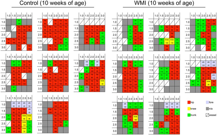
Hypoxia-ischemia (H-I) in rats at postnatal day 3 causes disorganization of oligodendrocyte development in layers II/III of the sensorimotor cortex without apparent neuronal loss, and shows mild hindlimb dysfunction with imbalanced motor coordination. However, the mechanisms by which mild motor dysfunction is induced without loss of cortical neurons are currently unclear. To reveal the mechanisms underlying mild motor dysfunction in neonatal H-I model, electrical responsiveness and dendrite morphology in the sensorimotor cortex were investigated at 10 weeks of age. Responses to intracortical microstimulation (ICMS) revealed that the cortical motor map was significantly changed in this model. The cortical area related to hip joint movement was reduced, and the area related to trunk movement was increased. Sholl analysis in Golgi staining revealed that layer I-III neurons on the H-I side had more dendrite branches compared with the contralateral side. To investigate whether changes in the motor map and morphology appeared at earlier stages, ICMS and Sholl analysis were also performed at 5 weeks of age. The minimal ICMS current to evoke twitches of the hip area was higher on the H-I side, while the motor map was unchanged. Golgi staining revealed more dendrite branches in layer I-III neurons on the H-I side. These results revealed that alterations of both dendrite morphology and ICMS threshold of the hip area occurred before the rearrangement of the motor map in the neonatal H-I model. They also suggest that altered dendritic morphology and altered ICMS responsiveness may be related to mild motor dysfunction in this model.
出生后第3天的大鼠缺氧缺血(H-I)会导致感觉运动皮层II/III层少突胶质细胞发育紊乱,而无明显神经元损失,并表现出轻度后肢功能障碍及运动协调失衡。然而,目前尚不清楚在无皮层神经元损失的情况下诱发轻度运动功能障碍的机制。为揭示新生儿H-I模型中轻度运动功能障碍的潜在机制,在10周龄时研究了感觉运动皮层的电反应性和树突形态。对皮层内微刺激(ICMS)的反应表明,该模型中的皮层运动图谱发生了显著变化。与髋关节运动相关的皮层区域减小,与躯干运动相关的区域增大。高尔基染色的Sholl分析显示,与对侧相比,H-I侧的I-III层神经元有更多的树突分支。为研究运动图谱和形态的变化是否在更早阶段出现,在5周龄时也进行了ICMS和Sholl分析。H-I侧诱发髋关节区域抽搐所需的最小ICMS电流更高,而运动图谱未改变。高尔基染色显示H-I侧的I-III层神经元有更多的树突分支。这些结果表明,在新生儿H-I模型中,树突形态和髋关节区域ICMS阈值的改变发生在运动图谱重新排列之前。它们还表明,树突形态改变和ICMS反应性改变可能与该模型中的轻度运动功能障碍有关。