Laboratory of Τumor Cell Biology, School of Medicine, University of Crete, Heraklion, Greece.
Department of Biochemistry, University of Crete, Greece Medical School, Heraklion, Greece.
Breast Cancer Res. 2018 Jul 5;20(1):67. doi: 10.1186/s13058-018-0993-z.
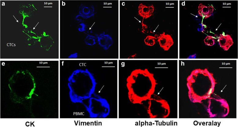
Circulating tumor cells (CTCs) are the major players in the metastatic process. A potential mechanism of cell migration and invasion is the formation of microtentacles in tumor cells. These structures are supported by α-tubulin (TUB), detyrosinated α-tubulin (GLU), and vimentin (VIM). In the current study, we evaluated the expression of those cytoskeletal proteins in CTCs.
Forty patients with breast cancer (BC) (16 early and 24 metastatic) were enrolled in the study. CTCs were isolated using the ISET platform and stained with the following combinations of antibodies: pancytokeratin (CK)/VIM/TUB and CK/VIM/GLU. Samples were analyzed with the ARIOL platform and confocal laser scanning microscopy.
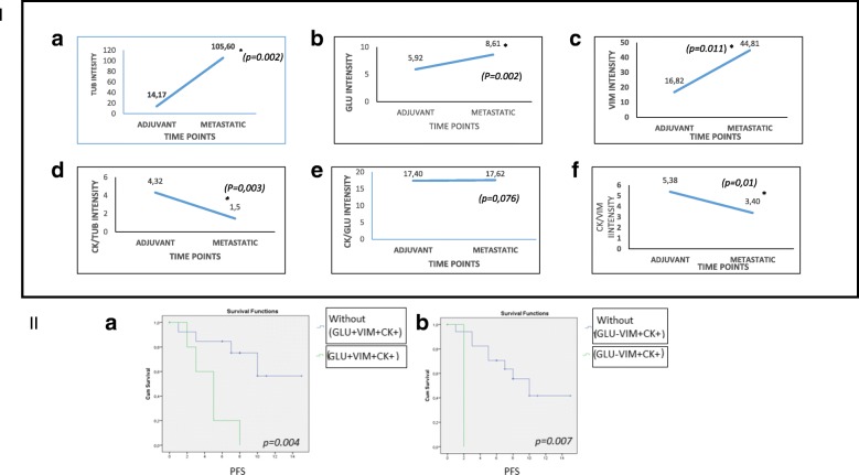
Fluorescence quantification revealed that the ratios CK/TUB, CK/VIM, and CK/GLU were statistically increased in MCF7 compared with more aggressive cell lines (SKBR3 and MDA-MB-231). In addition, all of these ratios were statistically increased in MCF7 cells compared with metastatic BC patients' CTCs (p = 0.0001, p = 0.0001, and p = 0.003, respectively). Interestingly, intercellular connections among CTCs and between CTCs and blood cells through cytoskeleton bridges were revealed, whereas microtentacles were increased in patients with CTC clusters. These intercellular connections were supported by TUB, VIM, and GLU. Quantification of the examined molecules revealed that the median intensity of TUB, GLU, and VIM was significantly increased in patients with metastatic BC compared with those with early disease (TUB, 62.27 vs 11.5, p = 0.0001; GLU, 6.99 vs 5.29, p = 0.029; and VIM, 8.24 vs 5.38, p = 0.0001, respectively).
CTCs from patients with BC aggregate to each other and to blood cells through cytoskeletal protrusions, supported by VIM, TUB, and GLU. Quantification of these molecules could potentially identify CTCs related to more aggressive disease.
循环肿瘤细胞(CTCs)是转移过程中的主要参与者。肿瘤细胞中微绒毛的形成是细胞迁移和侵袭的潜在机制之一。这些结构由α-微管蛋白(TUB)、去酪氨酸化的α-微管蛋白(GLU)和波形蛋白(VIM)支撑。在本研究中,我们评估了 CTC 中这些细胞骨架蛋白的表达。
本研究纳入了 40 名乳腺癌(BC)患者(16 名早期和 24 名转移性)。使用 ISET 平台分离 CTC ,并用以下抗体组合进行染色:细胞角蛋白(CK)/VIM/TUB 和 CK/VIM/GLU。使用 ARIOL 平台和共聚焦激光扫描显微镜对样本进行分析。
荧光定量显示,与更具侵袭性的细胞系(SKBR3 和 MDA-MB-231)相比,MCF7 中 CK/TUB、CK/VIM 和 CK/GLU 的比值均有统计学意义的增加。此外,与转移性 BC 患者的 CTC 相比,MCF7 细胞中所有这些比值均有统计学意义的增加(p=0.0001、p=0.0001 和 p=0.003)。有趣的是,在 CTC 簇的患者中,通过细胞骨架桥,在 CTC 之间以及 CTC 与血细胞之间显示出细胞间连接,并且微绒毛增加。这些细胞间连接由 TUB、VIM 和 GLU 支撑。对所检查分子的定量分析显示,与早期疾病患者相比,转移性 BC 患者的 TUB、GLU 和 VIM 的中位强度显著增加(TUB,62.27 与 11.5,p=0.0001;GLU,6.99 与 5.29,p=0.029;VIM,8.24 与 5.38,p=0.0001)。
BC 患者的 CTC 通过由 VIM、TUB 和 GLU 支撑的细胞骨架突起聚集在一起,与血液细胞聚集在一起。这些分子的定量分析可能有助于识别与侵袭性更强的疾病相关的 CTC。