Bae Sang Mun, Park Soo Jung, Choi Myoungeun, Song Miyeoun, Cho Young Eun, Do Eun-Ju, Ryu Yeon-Mi, Park Sunha, Lee Byung-Heon, Lee Sang-Wook, Hwang Sung Wook, Park Sang Hyoung, Yang Dong-Hoon, Ye Byong Duk, Byeon Jeong-Sik, Yang Suk-Kyun, Joo Jinmyoung, Kim Sang-Yeob, Myung Seung-Jae
Asan Institute for Life Science, Asan Medical Center, University of Ulsan College of medicine, Seoul, Republic of Korea.
Asan Institute for Life Science, Asan Medical Center, University of Ulsan College of medicine, Seoul, Republic of Korea; Department of Internal Medicine and Institute of Gastroenterology, Yonsei University College of medicine, Seoul, Republic of Korea.
Transl Oncol. 2018 Aug;11(4):1044-1052. doi: 10.1016/j.tranon.2018.06.008. Epub 2018 Jul 5.
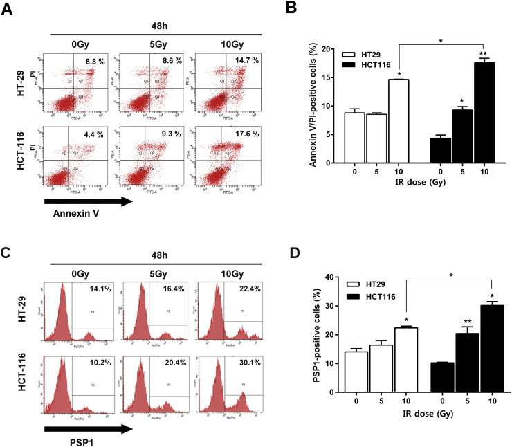
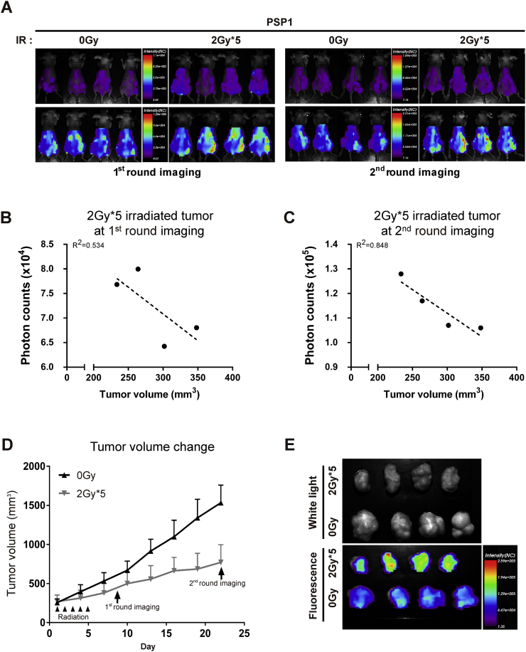
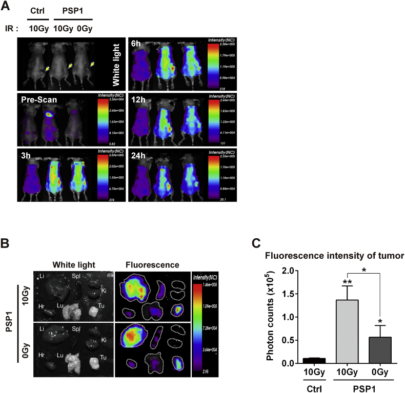
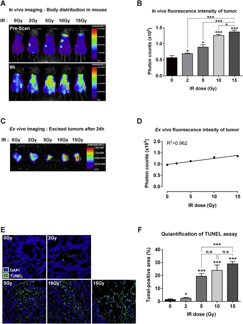
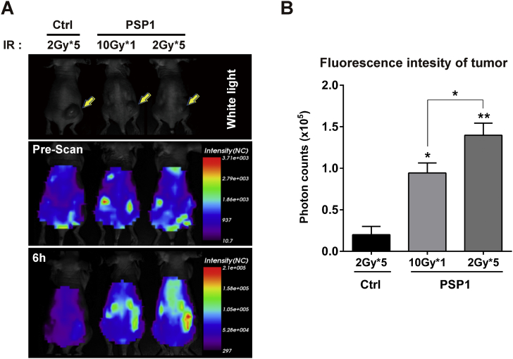
Accurate and timely visualization of apoptotic status in response to radiation is necessary for deciding whether to continue radiation or change to another mode of treatment. This is especially critical in patients with colorectal cancer, which requires a delicate combination of surgery, radiation, and chemotherapy in order to achieve optimal outcome. In this study, we investigated the potential of phosphatidylserine-recognizing peptide 1 (PSP1) as an apoptosis-targeting probe, which identifies phosphatidylserine on cell surfaces. We first screened colon cancer cell lines for their sensitivity to radiation and selected two cell lines: HCT116 and HT29. Cell binding assay using fluorescence-activated cell sorting and optical imaging showed that HCT116 cells had better binding to PSP1 than HT29 cells. Thus, mouse xenograft model using HCT116 cells was generated and was topically irradiated with either single or fractionated dose of radiation followed by systemic administration of PSP1 for subsequent molecular optical imaging. We confirmed that the PSP1 probe was selectively bound to apoptosis-induced tumor in a radiation dose-dependent manner. We also observed that fractionated radiation regimen, which is recently being used in clinical situation, was more effective in inducing tumor apoptosis than corresponding single-dose radiation treatment. We then evaluated the correlation between tumor targeting of PSP1 and suppression effect of tumor development and found that tumor volume and fluorescence intensity were correlated before (correlation coefficient r = 0.534) and after (r = 0.848) radiation therapy. Our study shows that PSP1 peptide is an efficient index probe for deciding "go or no-go" for radiation therapy in colorectal cancer.
准确及时地可视化辐射诱导的凋亡状态对于决定是否继续放疗或改用其他治疗方式至关重要。这在结直肠癌患者中尤为关键,因为结直肠癌需要手术、放疗和化疗的精细结合才能实现最佳治疗效果。在本研究中,我们研究了磷脂酰丝氨酸识别肽1(PSP1)作为一种靶向凋亡探针的潜力,该探针可识别细胞表面的磷脂酰丝氨酸。我们首先筛选了结肠癌细胞系对辐射的敏感性,并选择了两种细胞系:HCT116和HT29。使用荧光激活细胞分选和光学成像的细胞结合试验表明,HCT116细胞比HT29细胞与PSP1的结合更好。因此,构建了使用HCT116细胞的小鼠异种移植模型,对其进行单次或分次剂量的局部照射,随后全身给予PSP1进行后续的分子光学成像。我们证实,PSP1探针以辐射剂量依赖性方式选择性地结合到凋亡诱导的肿瘤上。我们还观察到,目前临床中使用的分次放疗方案在诱导肿瘤凋亡方面比相应的单次剂量放疗更有效。然后,我们评估了PSP1的肿瘤靶向性与肿瘤生长抑制效果之间的相关性,发现放疗前后肿瘤体积与荧光强度均具有相关性(放疗前相关系数r = 0.534,放疗后r = 0.848)。我们的研究表明,PSP1肽是决定结直肠癌放疗“继续或停止”的有效指标探针。