Department of Pharmacy, Health and Nutritional Sciences, University of Calabria, 87036 Rende, Italy.
Breast Cancer Now Research Unit, Division of Cancer Sciences, Manchester Cancer Research Centre, University of Manchester, Wilmslow Road, Manchester M20 4GJ, UK.
Int J Mol Sci. 2018 Jul 10;19(7):2011. doi: 10.3390/ijms19072011.
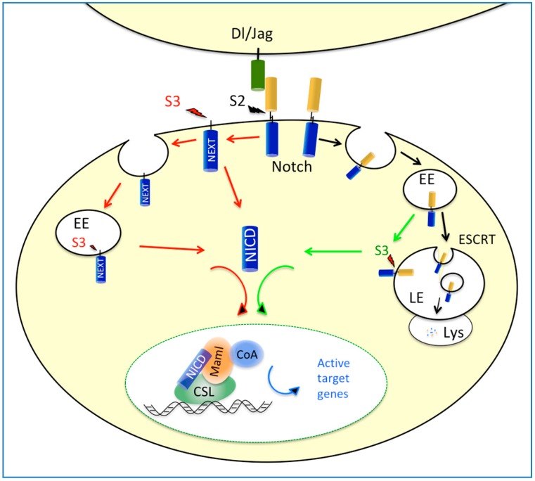
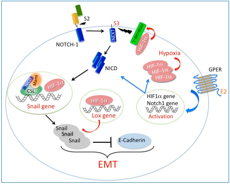
The Notch signaling pathway acts in both physiological and pathological conditions, including embryonic development and tumorigenesis. In cancer progression, diverse mechanisms are involved in Notch-mediated biological responses, including angiogenesis and epithelial-mesenchymal-transition (EMT). During EMT, the activation of cellular programs facilitated by transcriptional repressors results in epithelial cells losing their differentiated features, like cell–cell adhesion and apical–basal polarity, whereas they gain motility. As it concerns cancer epithelial cells, EMT may be consequent to the evolution of genetic/epigenetic instability, or triggered by factors that can act within the tumor microenvironment. Following a description of the Notch signaling pathway and its major regulatory nodes, we focus on studies that have given insights into the functional interaction between Notch signaling and either hypoxia or estrogen in breast cancer cells, with a particular focus on EMT. Furthermore, we describe the role of hypoxia signaling in breast cancer cells and discuss recent evidence regarding a functional interaction between HIF-1α and GPER in both breast cancer cells and cancer-associated fibroblasts (CAFs). On the basis of these studies, we propose that a functional network between HIF-1α, GPER and Notch may integrate tumor microenvironmental cues to induce robust EMT in cancer cells. Further investigations are required in order to better understand how hypoxia and estrogen signaling may converge on Notch-mediated EMT within the context of the stroma and tumor cells interaction. However, the data discussed here may anticipate the potential benefits of further pharmacological strategies targeting breast cancer progression.
Notch 信号通路在生理和病理条件下都有作用,包括胚胎发育和肿瘤发生。在癌症进展过程中,多种机制参与了 Notch 介导的生物学反应,包括血管生成和上皮间质转化 (EMT)。在 EMT 过程中,转录抑制剂激活细胞程序,导致上皮细胞失去分化特征,如细胞间黏附性和顶端-基底极性,而获得迁移能力。就癌症上皮细胞而言,EMT 可能是遗传/表观遗传不稳定性进化的结果,也可能是肿瘤微环境中某些因素触发的。在描述了 Notch 信号通路及其主要调控节点之后,我们重点研究了 Notch 信号与缺氧或雌激素在乳腺癌细胞中的功能相互作用的研究,特别关注 EMT。此外,我们还描述了缺氧信号在乳腺癌细胞中的作用,并讨论了最近关于 HIF-1α 和 GPER 在乳腺癌细胞和癌相关成纤维细胞 (CAFs) 中功能相互作用的证据。基于这些研究,我们提出 HIF-1α、GPER 和 Notch 之间的功能网络可能整合肿瘤微环境线索,诱导癌细胞发生强烈的 EMT。需要进一步的研究来更好地理解缺氧和雌激素信号如何在基质和肿瘤细胞相互作用的背景下汇集到 Notch 介导的 EMT 上。然而,这里讨论的数据可能预示着针对乳腺癌进展的进一步药理学策略的潜在益处。