Legin A A, Theiner S, Schintlmeister A, Reipert S, Heffeter P, Jakupec M A, Mayr J, Varbanov H P, Kowol C R, Galanski M, Berger W, Wagner M, Keppler B K
Institute of Inorganic Chemistry , Research Platform "Translational Cancer Therapy Research," and Research Network "Chemistry meets Microbiology" , University of Vienna , Währinger Straße 42 , A-1090 Vienna , Austria . Email:
Department of Microbiology and Ecosystem Science , Research Network "Chemistry meets Microbiology", and Large-Instrument Facility for Advanced Isotope Research , University of Vienna , A-1090 Vienna , Austria.
Chem Sci. 2016 May 1;7(5):3052-3061. doi: 10.1039/c5sc04383b. Epub 2016 Feb 3.
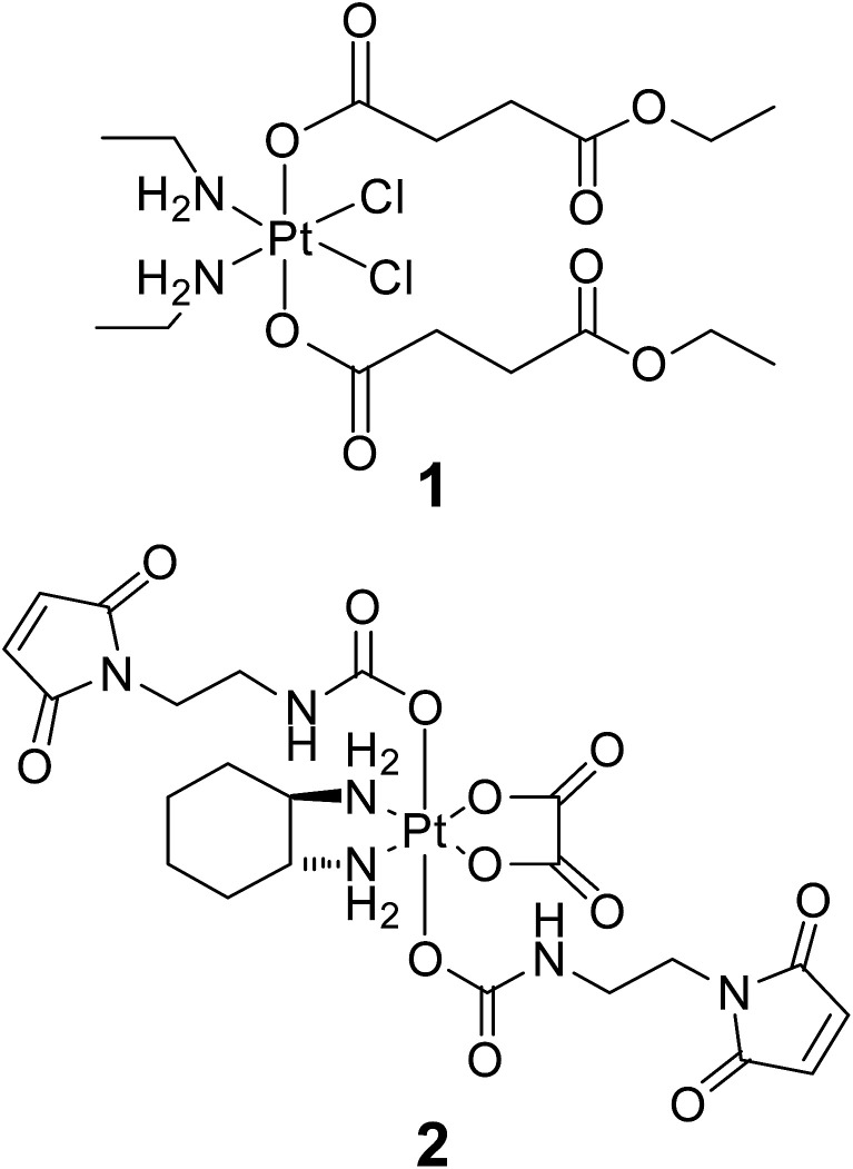
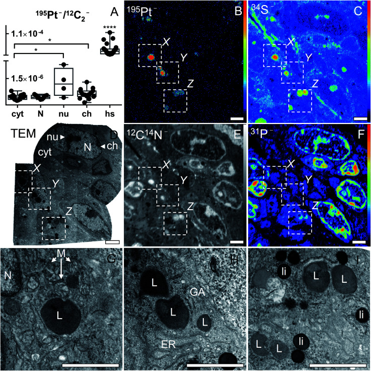
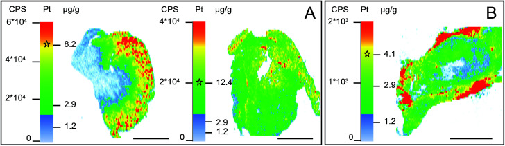
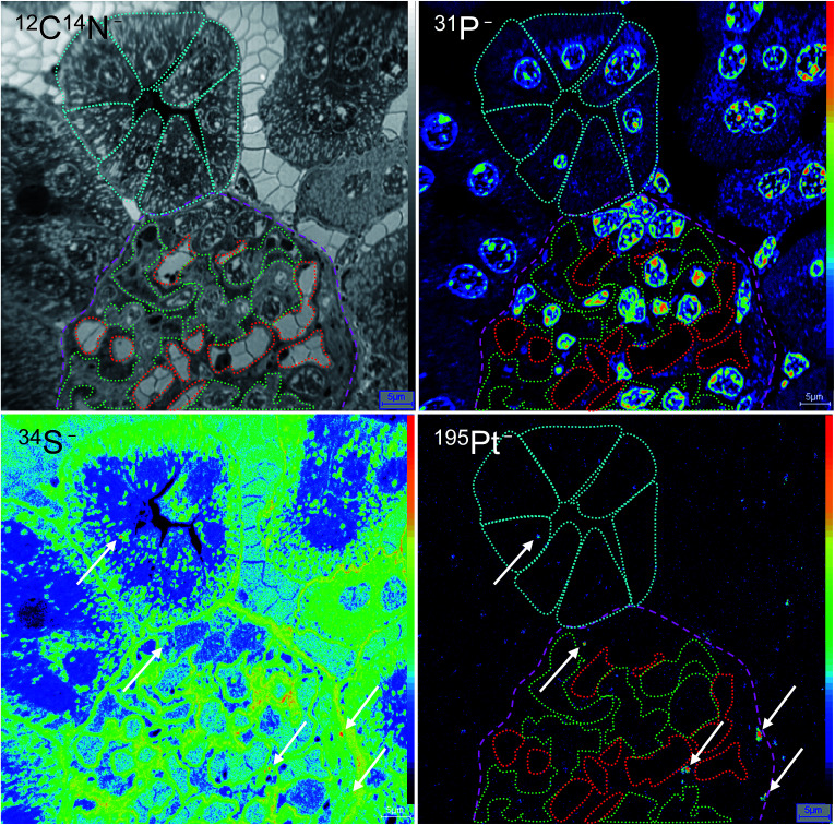
Nano-scale secondary ion mass spectrometry (NanoSIMS) enables trace element and isotope analyses with high spatial resolution. This unique capability has recently been exploited in several studies analyzing the subcellular distribution of Au and Pt anticancer compounds. However, these studies were restricted to cell culture systems. To explore the applicability to the setting, we developed a combined imaging approach consisting of laser ablation inductively coupled plasma mass spectrometry (LA-ICP-MS), NanoSIMS and transmission electron microscopy (TEM) suitable for multi-scale detection of the platinum distribution in tissues. Applying this approach to kidney and tumor samples upon administration of selected platinum(iv) anticancer prodrugs revealed uneven platinum distributions on both the organ and subcellular scales. Spatial platinum accumulation patterns were quantitatively assessed by LA-ICP-MS in histologically heterogeneous organs (, higher platinum accumulation in kidney cortex than in medulla) and used to select regions of interest for subcellular-scale imaging with NanoSIMS. These analyses revealed cytoplasmic sulfur-rich organelles accumulating platinum in both kidney and malignant cells. Those in the tumor were subsequently identified as organelles of lysosomal origin, demonstrating the potential of the combinatorial approach for investigating therapeutically relevant drug concentrations on a submicrometer scale.
纳米级二次离子质谱(NanoSIMS)能够实现具有高空间分辨率的微量元素和同位素分析。这种独特的能力最近在几项分析金和铂抗癌化合物亚细胞分布的研究中得到了应用。然而,这些研究仅限于细胞培养系统。为了探索其在体内环境中的适用性,我们开发了一种由激光烧蚀电感耦合等离子体质谱(LA-ICP-MS)、NanoSIMS和透射电子显微镜(TEM)组成的联合成像方法,适用于对组织中铂分布进行多尺度检测。将这种方法应用于给予选定的铂(IV)抗癌前药后的肾脏和肿瘤样本,发现在器官和亚细胞尺度上铂的分布均不均匀。通过LA-ICP-MS对组织学上异质的器官(如肾皮质中的铂积累高于髓质)中的铂积累模式进行定量评估,并用于选择感兴趣的区域进行NanoSIMS亚细胞尺度成像。这些分析揭示了富含硫的细胞质细胞器在肾脏和恶性细胞中都积累了铂。肿瘤中的这些细胞器随后被鉴定为溶酶体起源的细胞器,证明了这种组合方法在亚微米尺度上研究治疗相关药物浓度的潜力。