The State Key Laboratory Breeding Base of Basic Science of Stomatology (Hubei-MOST) and Key Laboratory of Oral Biomedicine Ministry of Education, School and Hospital of Stomatology, Wuhan University, 237 Luoyu Road, Wuhan, China.
Department of Oral and Maxillofacial Surgery, School and Hospital of Stomatology, Wuhan University, 237 Luoyu Road, Wuhan, China.
Biomed Res Int. 2018 May 29;2018:7537630. doi: 10.1155/2018/7537630. eCollection 2018.
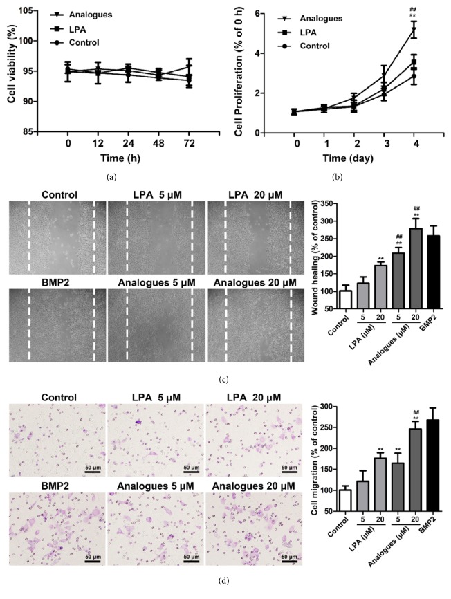
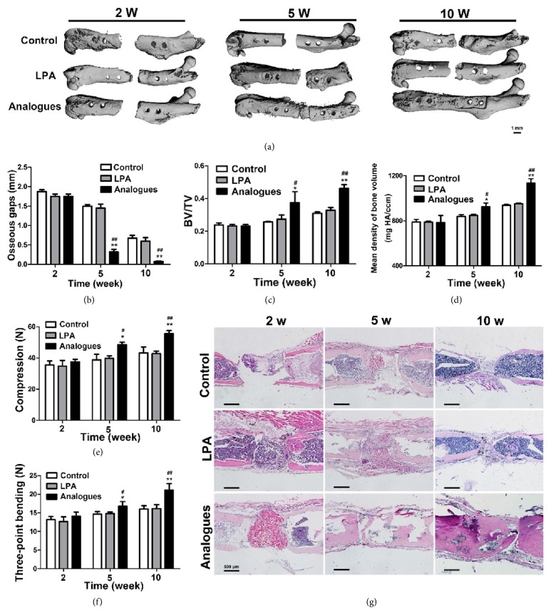
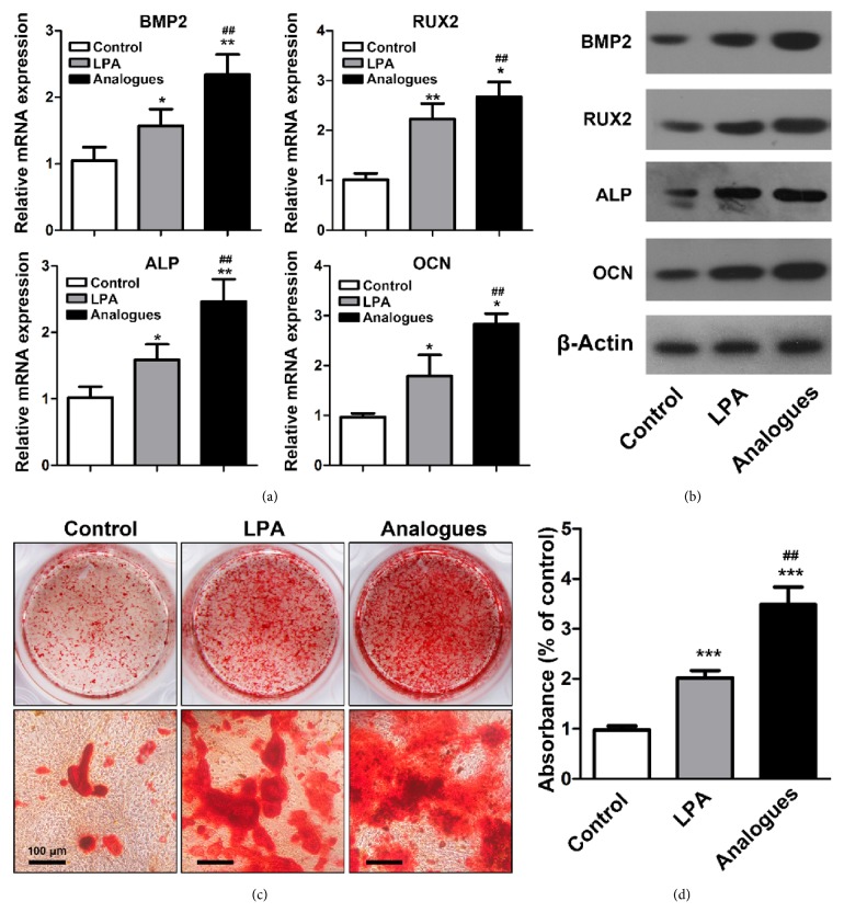
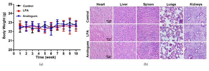
Lysophosphatidic acid (LPA), a bioactive lipid molecule, has recently emerged as physiological and pathophysiological regulator in skeletal biology. Here we evaluate the effects of LPA on bone formation in vivo in murine femoral critical defect model. Primary femoral osteoblasts were isolated and treated with osteogenic induction conditional media supplemented with 20 M LPA or LPA analogue. Mineralized nodules were visualized by Alizarin Red S staining. Forty-five C57BL/6 mice underwent unilateral osteotomy. The femoral osteotomy gap was filled with porous scaffolds of degradable chitosan/beta-tricalcium phosphate containing PBS, LPA, or LPA analogue. 2, 5, and 10 weeks after surgery, mice were sacrificed and femurs were harvested and prepared for Micro-Computed Tomography (Micro-CT) and histological analysis. Alizarin Red S staining showed that LPA and LPA analogue significantly enhanced the mineral deposition in osteoblasts. Micro-CT 3D reconstruction images and HE staining revealed that significantly more newly formed bone in osteotomy was treated with LPA analogue when compared to control and LPA group, which was verified by histological analysis and biomechanical characterization testing. In summary, our study demonstrated that although LPA promotes mineralized matrix formation in vitro, the locally administrated LPA was not effective in promoting bone formation in vivo. And bone formation was enhanced by LPA analogue, administrated locally in vivo. LPA analogue was a potent stimulating factor for bone formation in vivo due to its excellent stability.
溶血磷脂酸(LPA)是一种生物活性脂质分子,最近被认为是骨骼生物学中生理和病理生理学调节剂。在这里,我们评估了 LPA 对体内鼠股骨临界缺损模型中成骨的影响。分离原代股骨成骨细胞,用补充有 20μM LPA 或 LPA 类似物的成骨诱导条件培养基处理。通过茜素红 S 染色观察矿化结节。45 只 C57BL/6 小鼠接受单侧截骨术。多孔可降解壳聚糖/β-磷酸三钙支架的股骨截骨间隙用 PBS、LPA 或 LPA 类似物填充。手术后 2、5 和 10 周,处死小鼠,取出股骨并进行 Micro-CT(Micro-CT)和组织学分析。茜素红 S 染色显示 LPA 和 LPA 类似物显著增强了成骨细胞中的矿化沉积。Micro-CT 三维重建图像和 HE 染色显示,与对照组和 LPA 组相比,用 LPA 类似物处理的截骨处有更多的新骨形成,组织学分析和生物力学特征测试也证实了这一点。总之,我们的研究表明,尽管 LPA 促进了体外矿化基质的形成,但局部给予的 LPA 在体内并没有促进骨形成的作用。而 LPA 类似物则通过体内局部给药增强了骨形成。LPA 类似物由于其优异的稳定性,成为体内骨形成的有效刺激因子。