Program in Integrative Molecular and Biomedical Sciences, Baylor College of Medicine, Houston, TX, 77030-3411, USA.
Department of Pathology and Immunology, Baylor College of Medicine, Houston, TX, 77030-3411, USA.
Exp Eye Res. 2018 Nov;176:219-226. doi: 10.1016/j.exer.2018.07.014. Epub 2018 Jul 25.
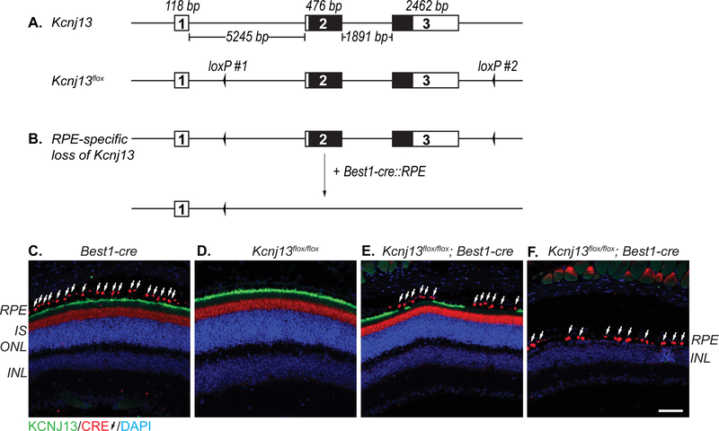
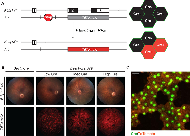
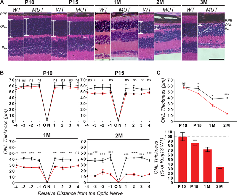
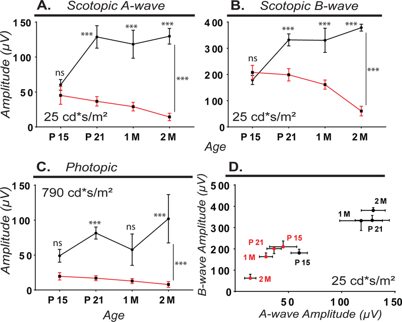
The retina is the light sensing tissue of the eye which contains multiple layers of cells required for the detection and transmission of a visual signal. Loss of the light-sensing photoreceptors leads to defects in visual function and blindness. Previously, we found that mosaic deletion of Kcnj13, and subsequent loss of the potassium channel Kir7.1, in mice leads to photoreceptor degeneration and recapitulates the human retinal disease phenotype (Zhong et al., 2015). Kcnj13 expression in the retinal pigment epithelium (RPE) is essential for normal retinal electrophysiology, function, and survival. Mice with homozygous loss of Kcnj13 die at postnatal day 1 (P1), requiring a tissue-specific approach to study retinal degeneration phenotypes in adult mice. We used the CRISPR-Cas9 system to generate a floxed, conditional loss-of-function (cKO) Kcnj13 allele to study the pathogenesis of Kcnj13 deficiency in the retina. To investigate if the Kcnj13 is required in the RPE for photoreceptor function and survival, we used Best1-cre, which is specifically expressed in the RPE. We observed complete loss of Kcnj13 expression in Cre-positive RPE cells. Furthermore, our findings show that widespread loss of Kcnj13 in the RPE leads to severe and progressive thinning of the outer nuclear layer and a reduced response to light. Finally, to detect Best1-cre expression in the RPE of live animals without sacrificing the animal for histology, we generated a Cre-reporter-containing Kcnj13 cKO mouse line (cKOR: Kcnj13; Best1-cre; Ai9) which can be rapidly screened using retinal fluorescence microscopy. These findings provide new tools for studying the roles of Kcnj13 in retinal homeostasis.
视网膜是眼睛的光感组织,包含多个细胞层,这些细胞层对于视觉信号的检测和传输是必需的。光感受器的丧失会导致视觉功能缺陷和失明。先前,我们发现小鼠中 Kcnj13 的镶嵌缺失以及随后的钾通道 Kir7.1 的缺失导致光感受器变性,并再现了人类视网膜疾病表型(Zhong 等人,2015 年)。视网膜色素上皮(RPE)中的 Kcnj13 表达对于正常的视网膜电生理学、功能和存活是必需的。Kcnj13 纯合缺失的小鼠在出生后第 1 天(P1)死亡,需要一种组织特异性方法来研究成年小鼠的视网膜变性表型。我们使用 CRISPR-Cas9 系统生成了一个 floxed、条件性功能丧失(cKO)Kcnj13 等位基因,以研究 Kcnj13 缺乏在视网膜中的发病机制。为了研究 Kcnj13 是否需要在 RPE 中维持光感受器的功能和存活,我们使用了特异性表达在 RPE 中的 Best1-cre。我们观察到 Cre 阳性 RPE 细胞中 Kcnj13 表达完全缺失。此外,我们的研究结果表明,RPE 中 Kcnj13 的广泛缺失导致外核层严重且进行性变薄以及对光的反应减弱。最后,为了在不牺牲动物进行组织学检查的情况下在活体动物的 RPE 中检测 Best1-cre 表达,我们生成了一种包含 Cre 报告基因的 Kcnj13 cKO 小鼠品系(cKOR:Kcnj13;Best1-cre;Ai9),可以通过视网膜荧光显微镜快速筛选。这些发现为研究 Kcnj13 在视网膜内稳态中的作用提供了新的工具。