• 文献检索
  • 文档翻译
  • 深度研究
  • 学术资讯
  • Suppr Zotero 插件Zotero 插件
  • 邀请有礼
  • 套餐&价格
  • 历史记录
应用&插件
Suppr Zotero 插件Zotero 插件浏览器插件Mac 客户端Windows 客户端微信小程序
定价
高级版会员购买积分包购买API积分包
服务
文献检索文档翻译深度研究API 文档MCP 服务
关于我们
关于 Suppr公司介绍联系我们用户协议隐私条款
关注我们

Suppr 超能文献

核心技术专利:CN118964589B侵权必究
粤ICP备2023148730 号-1Suppr @ 2026

文献检索

告别复杂PubMed语法,用中文像聊天一样搜索,搜遍4000万医学文献。AI智能推荐,让科研检索更轻松。

立即免费搜索

文件翻译

保留排版,准确专业,支持PDF/Word/PPT等文件格式,支持 12+语言互译。

免费翻译文档

深度研究

AI帮你快速写综述,25分钟生成高质量综述,智能提取关键信息,辅助科研写作。

立即免费体验

窖蛋白-3 基因敲除破坏小鼠心室肌细胞的 T 管结构并减少 T 管 I 密度。

Caveolin-3 KO disrupts t-tubule structure and decreases t-tubular I density in mouse ventricular myocytes.

机构信息

School of Physiology, Pharmacology and Neuroscience, Biomedical Sciences Building, University of Bristol , Bristol , United Kingdom.

Veterans Affairs San Diego Healthcare System and Department of Anesthesiology, University of California-San Diego , La Jolla, California.

出版信息

Am J Physiol Heart Circ Physiol. 2018 Nov 1;315(5):H1101-H1111. doi: 10.1152/ajpheart.00209.2018. Epub 2018 Jul 20.

DOI:10.1152/ajpheart.00209.2018
PMID:30028203
原文链接:https://pmc.ncbi.nlm.nih.gov/articles/PMC6415741/
Abstract

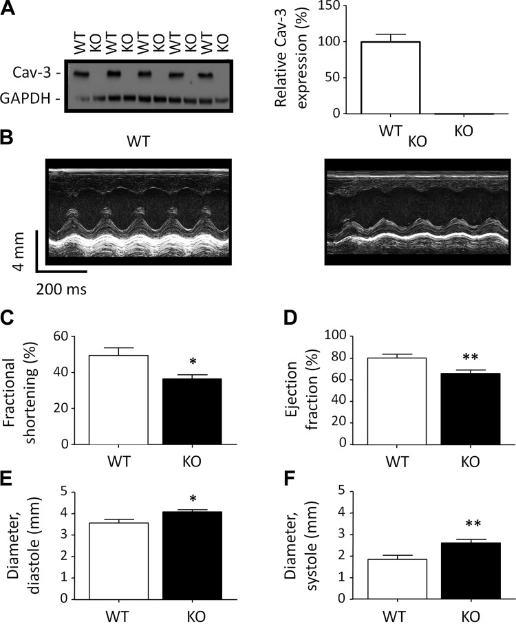
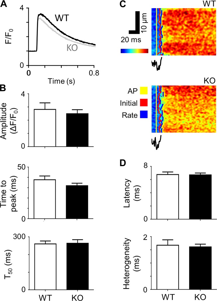
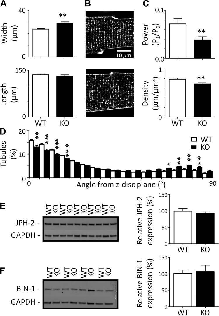
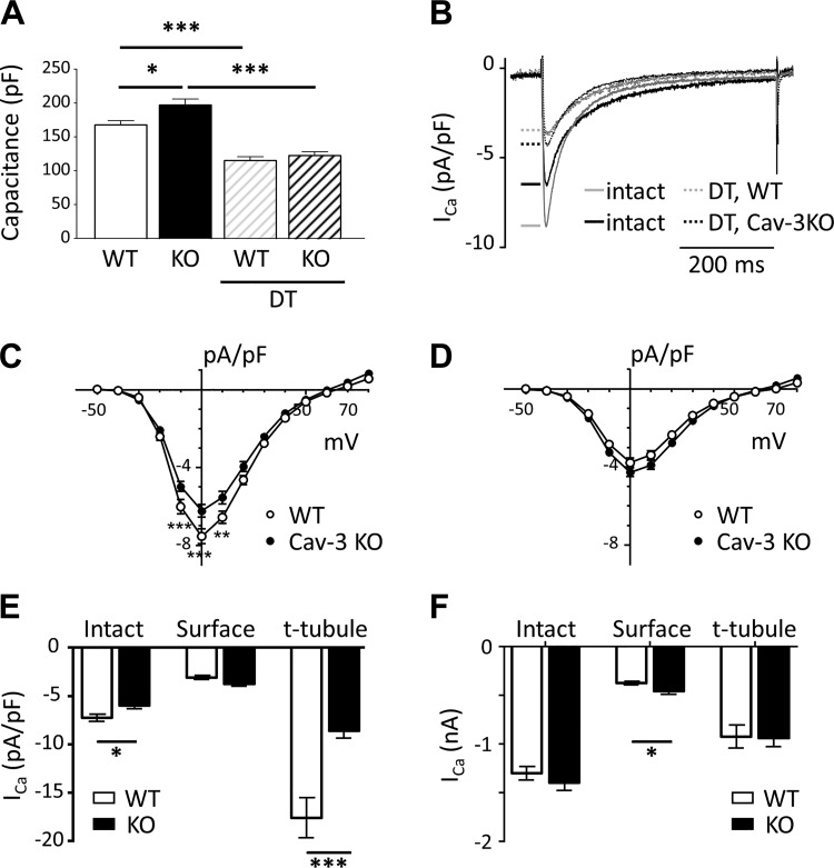
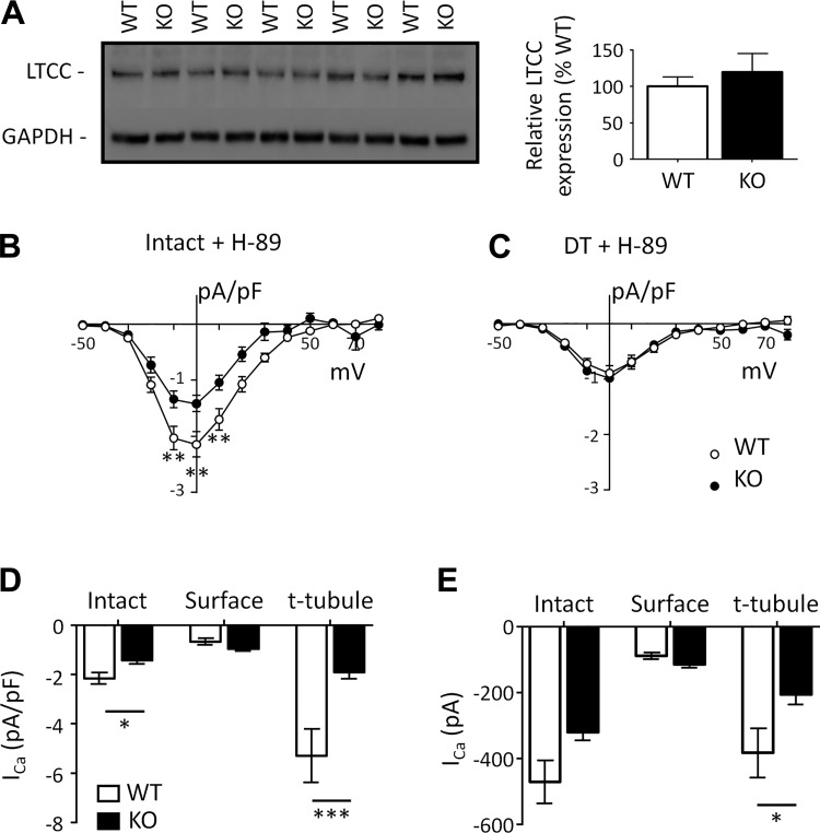
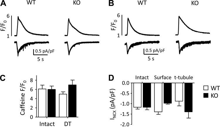
Caveolin-3 (Cav-3) is a protein that has been implicated in t-tubule formation and function in cardiac ventricular myocytes. In cardiac hypertrophy and failure, Cav-3 expression decreases, t-tubule structure is disrupted, and excitation-contraction coupling is impaired. However, the extent to which the decrease in Cav-3 expression underlies these changes is unclear. We therefore investigated the structure and function of myocytes isolated from the hearts of Cav-3 knockout (KO) mice. These mice showed cardiac dilatation and decreased ejection fraction in vivo compared with wild-type control mice. Isolated KO myocytes showed cellular hypertrophy, altered t-tubule structure, and decreased L-type Ca channel current ( I) density. This decrease in density occurred predominantly in the t-tubules, with no change in total I, and was therefore a consequence of the increase in membrane area. Cav-3 KO had no effect on L-type Ca channel expression, and C3SD peptide, which mimics the scaffolding domain of Cav-3, had no effect on I in KO myocytes. However, inhibition of PKA using H-89 decreased I at the surface and t-tubule membranes in both KO and wild-type myocytes. Cav-3 KO had no significant effect on Na/Ca exchanger current or Ca release. These data suggest that Cav-3 KO causes cellular hypertrophy, thereby decreasing t-tubular I density. NEW & NOTEWORTHY Caveolin-3 (Cav-3) is a protein that inhibits hypertrophic pathways, has been implicated in the formation and function of cardiac t-tubules, and shows decreased expression in heart failure. This study demonstrates that Cav-3 knockout mice show cardiac dysfunction in vivo, while isolated ventricular myocytes show cellular hypertrophy, changes in t-tubule structure, and decreased t-tubular L-type Ca current density, suggesting that decreased Cav-3 expression contributes to these changes in cardiac hypertrophy and failure.

摘要

窖蛋白-3(Cav-3)是一种蛋白,它与心肌细胞的 T 小管形成和功能有关。在心衰和衰竭中,Cav-3 的表达降低,T 小管结构破坏,兴奋-收缩偶联受损。然而,Cav-3 表达的降低在多大程度上导致了这些变化尚不清楚。因此,我们研究了 Cav-3 敲除(KO)小鼠心脏分离的心肌细胞的结构和功能。与野生型对照小鼠相比,这些小鼠在体内表现出心脏扩张和射血分数降低。分离的 KO 心肌细胞表现出细胞肥大、T 小管结构改变和 L 型钙通道电流(I)密度降低。这种密度的降低主要发生在 T 小管中,总 I 没有变化,因此是膜面积增加的结果。Cav-3 KO 对 L 型钙通道表达没有影响,C3SD 肽模拟 Cav-3 的支架结构域,对 KO 心肌细胞的 I 没有影响。然而,使用 H-89 抑制 PKA 会降低 KO 和野生型心肌细胞表面和 T 小管膜上的 I。Cav-3 KO 对 Na/Ca 交换器电流或 Ca 释放没有显著影响。这些数据表明,Cav-3 KO 导致细胞肥大,从而降低 T 小管 I 密度。新的和值得注意的是窖蛋白-3(Cav-3)是一种抑制肥大途径的蛋白,与心脏 T 小管的形成和功能有关,在心衰中表达降低。这项研究表明,Cav-3 KO 小鼠在体内表现出心脏功能障碍,而分离的心室肌细胞表现出细胞肥大、T 小管结构改变和 T 小管 L 型钙电流密度降低,表明 Cav-3 表达的降低导致了心脏肥大和衰竭中的这些变化。

https://cdn.ncbi.nlm.nih.gov/pmc/blobs/caf3/6415741/b568e6704809/zh40111826020006.jpg
https://cdn.ncbi.nlm.nih.gov/pmc/blobs/caf3/6415741/fcd0bf8d8b0a/zh40111826020001.jpg
https://cdn.ncbi.nlm.nih.gov/pmc/blobs/caf3/6415741/a2dcdfb65dfe/zh40111826020002.jpg
https://cdn.ncbi.nlm.nih.gov/pmc/blobs/caf3/6415741/ee43f450a481/zh40111826020003.jpg
https://cdn.ncbi.nlm.nih.gov/pmc/blobs/caf3/6415741/55ce43b28c39/zh40111826020004.jpg
https://cdn.ncbi.nlm.nih.gov/pmc/blobs/caf3/6415741/1d2f165bf253/zh40111826020005.jpg
https://cdn.ncbi.nlm.nih.gov/pmc/blobs/caf3/6415741/b568e6704809/zh40111826020006.jpg

相似文献

1
Caveolin-3 KO disrupts t-tubule structure and decreases t-tubular I density in mouse ventricular myocytes.窖蛋白-3 基因敲除破坏小鼠心室肌细胞的 T 管结构并减少 T 管 I 密度。
Am J Physiol Heart Circ Physiol. 2018 Nov 1;315(5):H1101-H1111. doi: 10.1152/ajpheart.00209.2018. Epub 2018 Jul 20.
2
Caveolin 3-dependent loss of t-tubular I during hypertrophy and heart failure in mice.小鼠肥大和心力衰竭过程中,小窝蛋白3依赖性横管肌浆网连接蛋白I的缺失
Exp Physiol. 2018 May 1;103(5):652-665. doi: 10.1113/EP086731. Epub 2018 Apr 14.
3
Cardiac-specific overexpression of caveolin-3 preserves t-tubular I during heart failure in mice.心肌特异性过表达窖蛋白-3可在小鼠心力衰竭过程中保持 T 管 I。
Exp Physiol. 2019 May;104(5):654-666. doi: 10.1113/EP087304. Epub 2019 Mar 14.
4
Altered distribution of ICa impairs Ca release at the t-tubules of ventricular myocytes from failing hearts.衰竭心脏心室肌细胞横管处ICa分布改变会损害钙释放。
J Mol Cell Cardiol. 2015 Sep;86:23-31. doi: 10.1016/j.yjmcc.2015.06.012. Epub 2015 Jun 21.
5
Loss of caveolin-3-dependent regulation of I in rat ventricular myocytes in heart failure.心力衰竭大鼠心室肌细胞中窖蛋白-3依赖的I调节丧失。
Am J Physiol Heart Circ Physiol. 2018 Mar 1;314(3):H521-H529. doi: 10.1152/ajpheart.00458.2017. Epub 2017 Nov 3.
6
The Effects of Aging on the Regulation of T-Tubular ICa by Caveolin in Mouse Ventricular Myocytes.衰老对小鼠心室肌细胞中 T 管钙电流调节的影响。
J Gerontol A Biol Sci Med Sci. 2018 May 9;73(6):711-719. doi: 10.1093/gerona/glx242.
7
Stimulation of ICa by basal PKA activity is facilitated by caveolin-3 in cardiac ventricular myocytes.基础 PKA 活性对 ICa 的刺激作用可被心肌细胞中的窖蛋白-3促进。
J Mol Cell Cardiol. 2014 Mar;68(100):47-55. doi: 10.1016/j.yjmcc.2013.12.026. Epub 2014 Jan 9.
8
Caveolin-3 Overexpression Attenuates Cardiac Hypertrophy via Inhibition of T-type Ca2+ Current Modulated by Protein Kinase Cα in Cardiomyocytes.小窝蛋白-3过表达通过抑制蛋白激酶Cα调控的心肌细胞T型钙电流减轻心肌肥厚
J Biol Chem. 2015 Sep 4;290(36):22085-100. doi: 10.1074/jbc.M115.674945. Epub 2015 Jul 13.
9
Interaction of the Joining Region in Junctophilin-2 With the L-Type Ca Channel Is Pivotal for Cardiac Dyad Assembly and Intracellular Ca Dynamics.衔接蛋白 Junctophilin-2 的连接区与 L 型钙通道的相互作用对于心肌二联体的组装和细胞内钙动力学至关重要。
Circ Res. 2021 Jan 8;128(1):92-114. doi: 10.1161/CIRCRESAHA.119.315715. Epub 2020 Oct 23.
10
Regulation of L-type calcium channel by phospholemman in cardiac myocytes.心肌细胞中磷蛋白对L型钙通道的调节作用。
J Mol Cell Cardiol. 2015 Jul;84:104-11. doi: 10.1016/j.yjmcc.2015.04.017. Epub 2015 Apr 25.

引用本文的文献

1
Ca microdomain-based excitation-transcription coupling in cardiac myocytes and vascular smooth muscle cells.心肌细胞和血管平滑肌细胞中基于钙微区的兴奋-转录偶联
Inflamm Regen. 2025 Jun 23;45(1):19. doi: 10.1186/s41232-025-00384-3.
2
Early Stage Morphogenesis of Transverse Tubules in Rat Cardiomyocytes: The Role of pBIN1.大鼠心肌细胞横小管的早期形态发生:pBIN1的作用
Circ Res. 2025 Jul 18;137(3):435-448. doi: 10.1161/CIRCRESAHA.124.325472. Epub 2025 Jun 13.
3
Ion Channel Regulation in Caveolae and Its Pathological Implications.

本文引用的文献

1
Caveolin 3-dependent loss of t-tubular I during hypertrophy and heart failure in mice.小鼠肥大和心力衰竭过程中,小窝蛋白3依赖性横管肌浆网连接蛋白I的缺失
Exp Physiol. 2018 May 1;103(5):652-665. doi: 10.1113/EP086731. Epub 2018 Apr 14.
2
The Effects of Aging on the Regulation of T-Tubular ICa by Caveolin in Mouse Ventricular Myocytes.衰老对小鼠心室肌细胞中 T 管钙电流调节的影响。
J Gerontol A Biol Sci Med Sci. 2018 May 9;73(6):711-719. doi: 10.1093/gerona/glx242.
3
Cholesterol depletion does not alter the capacitance or Ca handling of the surface or t-tubule membranes in mouse ventricular myocytes.
小窝中的离子通道调节及其病理意义。
Cells. 2025 Apr 24;14(9):631. doi: 10.3390/cells14090631.
4
Establishing a context of use for three-dimensional cardiac tissue derived from human-induced pluripotent stem cell-derived cardiomyocytes using inotropes.利用变力性药物建立源自人诱导多能干细胞衍生心肌细胞的三维心脏组织的使用背景。
Toxicol Sci. 2025 Jun 1;205(2):401-416. doi: 10.1093/toxsci/kfaf033.
5
Caveolin and oxidative stress in cardiac pathology.小窝蛋白与心脏病理学中的氧化应激
Front Physiol. 2025 Feb 18;16:1550647. doi: 10.3389/fphys.2025.1550647. eCollection 2025.
6
Rad protein: An essential player in L-type Ca2+ channel localization and modulation in cardiomyocytes.Rad 蛋白:在心肌细胞中 L 型 Ca2+通道定位和调节中的必需分子。
J Gen Physiol. 2024 Oct 7;156(10). doi: 10.1085/jgp.202413629. Epub 2024 Aug 22.
7
MG53/TRIM72: multi-organ repair protein and beyond.MG53/TRIM72:多器官修复蛋白及其他功能
Front Physiol. 2024 Apr 12;15:1377025. doi: 10.3389/fphys.2024.1377025. eCollection 2024.
8
Ptpn23 Controls Cardiac T-Tubule Patterning by Promoting the Assembly of Dystrophin-Glycoprotein Complex.Ptpn23通过促进肌营养不良蛋白-糖蛋白复合物的组装来控制心脏T小管的形态。
Circulation. 2024 Apr 23;149(17):1375-1390. doi: 10.1161/CIRCULATIONAHA.123.065767. Epub 2024 Jan 12.
9
Advances in the Study of MG53 in Cardiovascular Disease.MG53在心血管疾病中的研究进展
Int J Gen Med. 2023 Dec 23;16:6073-6082. doi: 10.2147/IJGM.S435030. eCollection 2023.
10
Unraveling the Cave: A Seventy-Year Journey into the Caveolar Network, Cellular Signaling, and Human Disease.揭开洞穴的奥秘:七十载探索陷窝网络、细胞信号转导与人类疾病
Cells. 2023 Nov 22;12(23):2680. doi: 10.3390/cells12232680.
胆固醇耗竭不会改变小鼠心室肌细胞表面或横管膜的电容或钙处理能力。
Physiol Rep. 2017 Nov;5(22). doi: 10.14814/phy2.13500.
4
Loss of caveolin-3-dependent regulation of I in rat ventricular myocytes in heart failure.心力衰竭大鼠心室肌细胞中窖蛋白-3依赖的I调节丧失。
Am J Physiol Heart Circ Physiol. 2018 Mar 1;314(3):H521-H529. doi: 10.1152/ajpheart.00458.2017. Epub 2017 Nov 3.
5
Sarcolemmal distribution of and and Ca autoregulation in mouse ventricular myocytes.小鼠心室肌细胞中 和 的肌膜分布及钙自动调节
Am J Physiol Heart Circ Physiol. 2017 Jul 1;313(1):H190-H199. doi: 10.1152/ajpheart.00117.2017. Epub 2017 May 5.
6
Junctophilin-2 gene therapy rescues heart failure by normalizing RyR2-mediated Ca release.连接蛋白-2基因疗法通过使兰尼碱受体2介导的钙释放正常化来挽救心力衰竭。
Int J Cardiol. 2016 Dec 15;225:371-380. doi: 10.1016/j.ijcard.2016.10.021. Epub 2016 Oct 8.
7
Elevated ventricular wall stress disrupts cardiomyocyte t-tubule structure and calcium homeostasis.升高的心室壁应力会破坏心肌细胞的横管结构和钙稳态。
Cardiovasc Res. 2016 Oct;112(1):443-51. doi: 10.1093/cvr/cvw111. Epub 2016 May 25.
8
Altered distribution of ICa impairs Ca release at the t-tubules of ventricular myocytes from failing hearts.衰竭心脏心室肌细胞横管处ICa分布改变会损害钙释放。
J Mol Cell Cardiol. 2015 Sep;86:23-31. doi: 10.1016/j.yjmcc.2015.06.012. Epub 2015 Jun 21.
9
Dependence of cardiac transverse tubules on the BAR domain protein amphiphysin II (BIN-1).心脏横小管对BAR结构域蛋白发动蛋白II(BIN-1)的依赖性。
Circ Res. 2014 Dec 5;115(12):986-96. doi: 10.1161/CIRCRESAHA.116.303448. Epub 2014 Oct 20.
10
Stimulation of ICa by basal PKA activity is facilitated by caveolin-3 in cardiac ventricular myocytes.基础 PKA 活性对 ICa 的刺激作用可被心肌细胞中的窖蛋白-3促进。
J Mol Cell Cardiol. 2014 Mar;68(100):47-55. doi: 10.1016/j.yjmcc.2013.12.026. Epub 2014 Jan 9.