Sullivan Travis B, Robert Litchfield C, Teebagy Patrick A, Morgan Shannon E, Beatty Evan W, Cicuto Bryan J, Nowd Peter K, Rieger-Christ Kimberly M, Bryan David J
Department of Translational Research, Lahey Hospital & Medical Center, Burlington, MA, USA.
Tissue Engineering Laboratory, Lahey Hospital & Medical Center, Burlington, MA, USA.
Neural Regen Res. 2018 Jul;13(7):1253-1262. doi: 10.4103/1673-5374.235073.
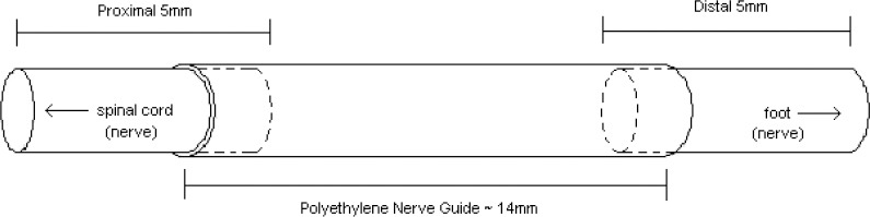
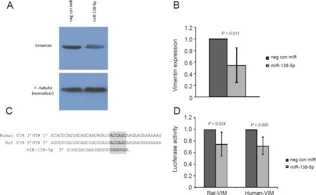
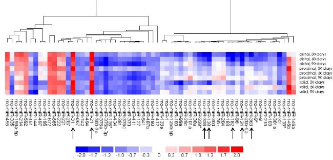
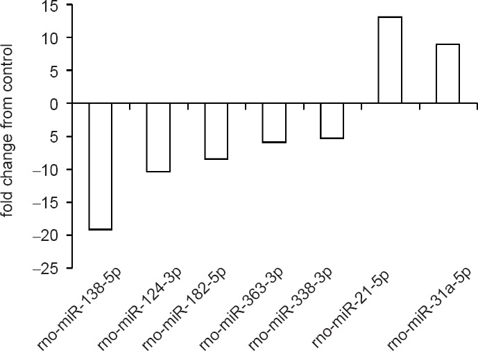
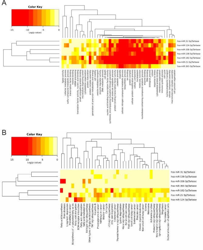
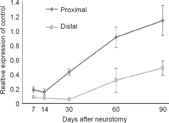
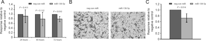
While the peripheral nervous system has regenerative ability, restoration of sufficient function remains a challenge. Vimentin has been shown to be localized in axonal growth fronts and associated with nerve regeneration, including myelination, neuroplasticity, kinase signaling in nerve axoplasm, and cell migration; however, the mechanisms regulating its expression within Schwann cell (SC) remain unexplored. The aim of this study was to profile the spatial and temporal expression profile of microRNA (miRNA) in a regenerating rat sciatic nerve after transection, and explore the potential role of miR-138-5p targeting vimentin in SC proliferation and migration. A rat sciatic nerve transection model, utilizing a polyethylene nerve guide, was used to investigate miRNA expression at 7, 14, 30, 60, and 90 days during nerve regeneration. Relative levels of miRNA expression were determined using microarray analysis and subsequently validated with quantitative real-time polymerase chain reaction. In vitro assays were conducted with cultured Schwann cells transfected with miRNA mimics and assessed for migratory and proliferative potential. The top seven dysregulated miRNAs reported in this study have been implicated in cell migration elsewhere, and GO and KEGG analyses predicted activities essential to wound healing. Transfection of one of these, miRNA-138-5p, into SCs reduced cell migration and proliferation. miR-138-5p has been shown to directly target vimentin in cancer cells, and the luciferase assay performed here in rat Schwann cells confirmed it. These results detail a role of miR-138-5p in rat peripheral nerve regeneration and expand on reports of it as an important regulator in the peripheral nervous system.
虽然外周神经系统具有再生能力,但恢复足够的功能仍然是一项挑战。波形蛋白已被证明定位于轴突生长前沿,并与神经再生相关,包括髓鞘形成、神经可塑性、神经轴浆中的激酶信号传导和细胞迁移;然而,调节其在施万细胞(SC)内表达的机制仍未被探索。本研究的目的是剖析大鼠坐骨神经横断后再生过程中微小RNA(miRNA)的时空表达谱,并探讨miR-138-5p靶向波形蛋白在施万细胞增殖和迁移中的潜在作用。利用聚乙烯神经导管的大鼠坐骨神经横断模型,用于研究神经再生过程中第7、14、30、60和90天的miRNA表达。使用微阵列分析确定miRNA表达的相对水平,随后用定量实时聚合酶链反应进行验证。对用miRNA模拟物转染的培养施万细胞进行体外试验,并评估其迁移和增殖潜力。本研究报告的前七种失调miRNA与其他地方的细胞迁移有关,GO和KEGG分析预测了对伤口愈合至关重要的活性。将其中一种miRNA-138-5p转染到施万细胞中可降低细胞迁移和增殖。miR-138-5p已被证明在癌细胞中直接靶向波形蛋白,此处对大鼠施万细胞进行的荧光素酶测定证实了这一点。这些结果详细阐述了miR-138-5p在大鼠周围神经再生中的作用,并扩展了其作为周围神经系统重要调节因子的报道。