• 文献检索
  • 文档翻译
  • 深度研究
  • 学术资讯
  • Suppr Zotero 插件Zotero 插件
  • 邀请有礼
  • 套餐&价格
  • 历史记录
应用&插件
Suppr Zotero 插件Zotero 插件浏览器插件Mac 客户端Windows 客户端微信小程序
定价
高级版会员购买积分包购买API积分包
服务
文献检索文档翻译深度研究API 文档MCP 服务
关于我们
关于 Suppr公司介绍联系我们用户协议隐私条款
关注我们

Suppr 超能文献

核心技术专利:CN118964589B侵权必究
粤ICP备2023148730 号-1Suppr @ 2026

文献检索

告别复杂PubMed语法,用中文像聊天一样搜索,搜遍4000万医学文献。AI智能推荐,让科研检索更轻松。

立即免费搜索

文件翻译

保留排版,准确专业,支持PDF/Word/PPT等文件格式,支持 12+语言互译。

免费翻译文档

深度研究

AI帮你快速写综述,25分钟生成高质量综述,智能提取关键信息,辅助科研写作。

立即免费体验

通过对活检标本进行颜色编码以降低根治性前列腺切除术中的阳性切缘率,从而实现个体化保留神经血管束:这是否可行?一项初步研究。

Lowering positive margin rates at radical prostatectomy by color coding of biopsy specimens to permit individualized preservation of the neurovascular bundles: is it feasible? a pilot investigation.

机构信息

Department of Urology, Rush University Medical Center, Chicago, IL, USA.

出版信息

Int Braz J Urol. 2018 Nov-Dec;44(6):1081-1088. doi: 10.1590/S1677-5538.IBJU.2017.0328.

DOI:10.1590/S1677-5538.IBJU.2017.0328
PMID:30044594
原文链接:https://pmc.ncbi.nlm.nih.gov/articles/PMC6442172/
Abstract

OBJECTIVE

To evaluate whether color-coding of prostate core biopsy specimens aids in preservation of the neurovascular bundles from an oncological perspective.

MATERIALS AND METHODS

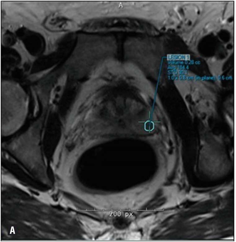
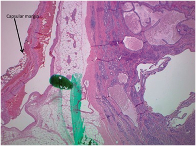
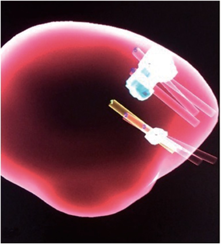
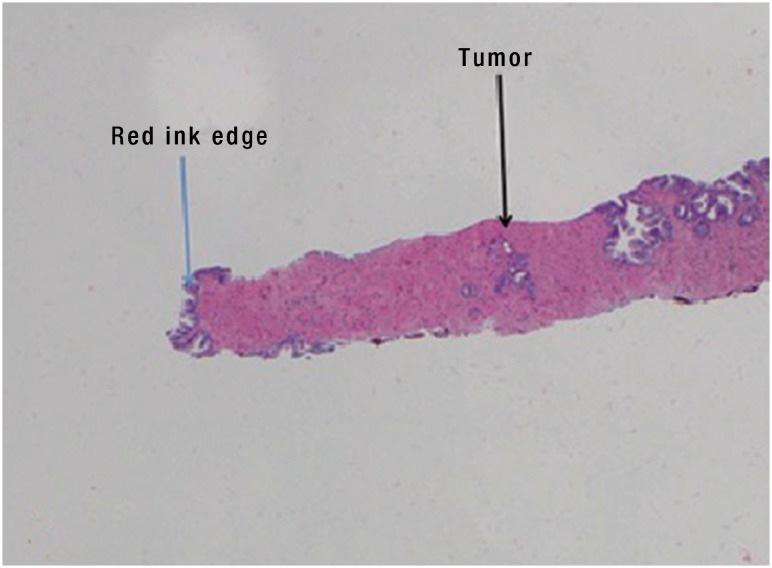
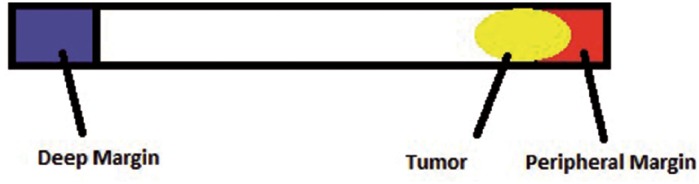
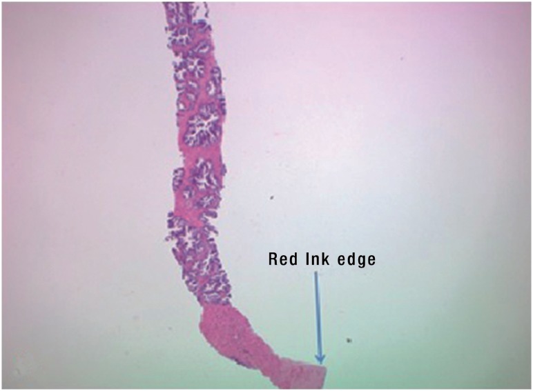
MRI guided transrectal ultrasound and biopsy of the prostate were performed in 51 consecutive patients suspected of being at high risk for harboring prostate cancer. Core specimens were labeled with blue dye at the deep aspect and red dye at the superficial peripheral aspect of the core. The distance from the tumor to the end of the dyed specimen was measured to determine if there was an area of normal tissue between the prostate capsule and tumor.

RESULTS

Of the 51 patients undergoing prostate biopsy, 30 (58.8%) were found to have cancer of the prostate: grade group 1 in 13.7%, 2 in 25.5%, 3 in 7.8%, 4 in 7.8% and 5 in 3.9% of the cohort. A total of 461 cores were analyzed in the cohort, of which 122 showed cancer. Five patients opted to undergo robotic assisted laparoscopic radical prostatectomy. No patients had a positive surgical margin (PSM) or extra prostatic extension (EPE) on radical prostatectomy if there was a margin of normal prostatic tissue seen between the dye and the tumor on prostate biopsy.

CONCLUSION

Color-coding of prostate biopsy core specimens may assist in tailoring the approach for preservation of the neurovascular bundles without compromising early oncological efficacy. Further study is required to determine whether this simple modification of the prostate biopsy protocol is valuable in larger groups of patients.

摘要

目的

评估前列腺核心活检标本的颜色编码是否有助于从肿瘤学角度保护神经血管束。

材料与方法

对 51 例疑似前列腺癌高危患者进行 MRI 引导经直肠超声和前列腺活检。核心标本在深部用蓝色染料标记,在核心的浅表周边用红色染料标记。测量从肿瘤到染色标本末端的距离,以确定前列腺包膜和肿瘤之间是否有正常组织区域。

结果

在接受前列腺活检的 51 例患者中,30 例(58.8%)发现前列腺癌:13.7%为 1 级,25.5%为 2 级,7.8%为 3 级,7.8%为 4 级,3.9%为 5 级。在该队列中分析了 461 个核心,其中 122 个显示癌症。5 名患者选择接受机器人辅助腹腔镜根治性前列腺切除术。如果在前列腺活检中,染料和肿瘤之间的前列腺组织有正常组织边缘,那么没有患者在根治性前列腺切除术中出现阳性手术切缘(PSM)或额外的前列腺外延伸(EPE)。

结论

前列腺活检核心标本的颜色编码可能有助于在不影响早期肿瘤疗效的情况下,针对神经血管束的保护调整方法。需要进一步研究以确定这种前列腺活检方案的简单修改是否在更大的患者群体中具有价值。

https://cdn.ncbi.nlm.nih.gov/pmc/blobs/5182/6442172/0fd2bfff231d/1677-6119-ibju-44-06-1081-gf06.jpg
https://cdn.ncbi.nlm.nih.gov/pmc/blobs/5182/6442172/2e43ddd88310/1677-6119-ibju-44-06-1081-gf01.jpg
https://cdn.ncbi.nlm.nih.gov/pmc/blobs/5182/6442172/05c5dc9cf28c/1677-6119-ibju-44-06-1081-gf02.jpg
https://cdn.ncbi.nlm.nih.gov/pmc/blobs/5182/6442172/0c2a27ab9e02/1677-6119-ibju-44-06-1081-gf03.jpg
https://cdn.ncbi.nlm.nih.gov/pmc/blobs/5182/6442172/336225ec0553/1677-6119-ibju-44-06-1081-gf04.jpg
https://cdn.ncbi.nlm.nih.gov/pmc/blobs/5182/6442172/d3ac023223f2/1677-6119-ibju-44-06-1081-gf05.jpg
https://cdn.ncbi.nlm.nih.gov/pmc/blobs/5182/6442172/0fd2bfff231d/1677-6119-ibju-44-06-1081-gf06.jpg

相似文献

1
Lowering positive margin rates at radical prostatectomy by color coding of biopsy specimens to permit individualized preservation of the neurovascular bundles: is it feasible? a pilot investigation.通过对活检标本进行颜色编码以降低根治性前列腺切除术中的阳性切缘率,从而实现个体化保留神经血管束:这是否可行?一项初步研究。
Int Braz J Urol. 2018 Nov-Dec;44(6):1081-1088. doi: 10.1590/S1677-5538.IBJU.2017.0328.
2
Focused Submission of Tissue for Radical Prostatectomy Following Multiparametric Magnetic Resonance Imaging/Ultrasound Fusion-Targeted Biopsy.多参数磁共振成像/超声融合靶向活检后针对根治性前列腺切除术的组织重点提交
Int J Surg Pathol. 2020 Feb;28(1):44-50. doi: 10.1177/1066896919865026. Epub 2019 Jul 25.
3
Magnetic resonance imaging-transectal ultrasound image-fusion biopsies accurately characterize the index tumor: correlation with step-sectioned radical prostatectomy specimens in 135 patients.磁共振成像-经直肠超声图像融合活检能准确描绘出目标肿瘤:与 135 例患者的分段式根治性前列腺切除术标本的相关性。
Eur Urol. 2015 Apr;67(4):787-94. doi: 10.1016/j.eururo.2014.08.077. Epub 2014 Sep 17.
4
Discordance between location of positive cores in biopsy and location of positive surgical margin following radical prostatectomy.前列腺穿刺活检阳性癌灶位置与根治性前列腺切除术后手术切缘阳性位置之间的不一致。
Korean J Urol. 2015 Oct;56(10):710-6. doi: 10.4111/kju.2015.56.10.710. Epub 2015 Oct 6.
5
Perineural invasion status, Gleason score and number of positive cores in biopsy pathology are predictors of positive surgical margin following laparoscopic radical prostatectomy.神经周围侵犯状态、Gleason评分以及活检病理中阳性核心的数量是腹腔镜根治性前列腺切除术后手术切缘阳性的预测指标。
Asian J Androl. 2017 Jul-Aug;19(4):468-472. doi: 10.4103/1008-682X.173444.
6
A comparison of the incidence and location of positive surgical margins in robotic assisted laparoscopic radical prostatectomy and open retropubic radical prostatectomy.机器人辅助腹腔镜根治性前列腺切除术与开放性耻骨后根治性前列腺切除术中阳性手术切缘的发生率及位置比较。
J Urol. 2007 Dec;178(6):2385-9; discussion 2389-90. doi: 10.1016/j.juro.2007.08.008. Epub 2007 Oct 22.
7
Pre-biopsy 3-Tesla MRI and targeted biopsy of the index prostate cancer: correlation with robot-assisted radical prostatectomy.活检前3特斯拉磁共振成像与索引前列腺癌的靶向活检:与机器人辅助根治性前列腺切除术的相关性
BJU Int. 2017 Jan;119(1):82-90. doi: 10.1111/bju.13525. Epub 2016 Jun 3.
8
Influence of magnetic resonance imaging in the decision to preserve or resect neurovascular bundles at robotic assisted laparoscopic radical prostatectomy.磁共振成像对机器人辅助腹腔镜根治性前列腺切除术中保留或切除神经血管束的决策的影响。
J Urol. 2014 Jul;192(1):82-8. doi: 10.1016/j.juro.2014.01.005. Epub 2014 Jan 15.
9
Higher number of transrectal ultrasound guided prostate biopsy cores is associated with higher blood loss and perioperative complications in robot assisted radical prostatectomy.在机器人辅助根治性前列腺切除术中,经直肠超声引导下前列腺穿刺活检芯数量较多与出血量增加及围手术期并发症相关。
Actas Urol Esp. 2017 Apr;41(3):155-161. doi: 10.1016/j.acuro.2016.09.012. Epub 2016 Nov 24.
10
The New York University nerve sparing algorithm decreases the rate of positive surgical margins following radical retropubic prostatectomy.纽约大学的神经保留算法降低了耻骨后根治性前列腺切除术后手术切缘阳性率。
J Urol. 2003 Jun;169(6):2147-52. doi: 10.1097/01.ju.0000057496.49676.5a.

引用本文的文献

1
Age and Body Mass Index: the most important factors of urinary and erectile function recovery after robotic assisted radical prostatectomy.年龄和体重指数:机器人辅助根治性前列腺切除术后排尿和勃起功能恢复的最重要因素。
Int Braz J Urol. 2019 Jul-Aug;45(4):653-654. doi: 10.1590/S1677-5538.IBJU.2019.04.01.

本文引用的文献

1
Central zone lesions on magnetic resonance imaging: Should we be concerned?磁共振成像上的中央区病变:我们应该担心吗?
Urol Oncol. 2017 Jan;35(1):31.e7-31.e12. doi: 10.1016/j.urolonc.2016.08.006. Epub 2016 Sep 28.
2
Periprostatic Fat: A Risk Factor for Prostate Cancer?前列腺周围脂肪:前列腺癌的一个风险因素?
Urology. 2016 Dec;98:107-112. doi: 10.1016/j.urology.2016.07.042. Epub 2016 Sep 1.
3
Missing the Mark: Prostate Cancer Upgrading by Systematic Biopsy over Magnetic Resonance Imaging/Transrectal Ultrasound Fusion Biopsy.
错失良机:系统活检通过磁共振成像/经直肠超声融合活检对前列腺癌的升级。
J Urol. 2017 Feb;197(2):327-334. doi: 10.1016/j.juro.2016.08.097. Epub 2016 Aug 28.
4
Tumor contact with prostate capsule on magnetic resonance imaging: A potential biomarker for staging and prognosis.磁共振成像上肿瘤与前列腺包膜的接触:一种用于分期和预后的潜在生物标志物。
Urol Oncol. 2017 Jan;35(1):30.e1-30.e8. doi: 10.1016/j.urolonc.2016.07.013. Epub 2016 Aug 24.
5
The concordance between the volume hotspot and the grade hotspot: a 3-D reconstructive model using the pathology outputs from the PROMIS trial.体积热点与分级热点之间的一致性:使用PROMIS试验病理结果的三维重建模型。
Prostate Cancer Prostatic Dis. 2016 Sep;19(3):258-63. doi: 10.1038/pcan.2016.7. Epub 2016 Jul 12.
6
The significance of anterior prostate lesions on multiparametric magnetic resonance imaging in African-American men.非洲裔美国男性前列腺前部病变在多参数磁共振成像中的意义。
Urol Oncol. 2016 Jun;34(6):254.e15-21. doi: 10.1016/j.urolonc.2015.12.018. Epub 2016 Feb 20.
7
Cancer statistics, 2016.癌症统计数据,2016 年。
CA Cancer J Clin. 2016 Jan-Feb;66(1):7-30. doi: 10.3322/caac.21332. Epub 2016 Jan 7.
8
Intraoperative Optical Biopsy during Robotic Assisted Radical Prostatectomy Using Confocal Endomicroscopy.术中使用共聚焦内窥镜行机器人辅助根治性前列腺切除术的光学活组织检查。
J Urol. 2016 Apr;195(4 Pt 1):1110-1117. doi: 10.1016/j.juro.2015.10.182. Epub 2015 Nov 26.
9
Updated prostate imaging reporting and data system (PIRADS v2) recommendations for the detection of clinically significant prostate cancer using multiparametric MRI: critical evaluation using whole-mount pathology as standard of reference.使用多参数磁共振成像检测临床显著性前列腺癌的更新版前列腺影像报告和数据系统(PIRADS v2)推荐意见:以全层病理作为参考标准的批判性评估
Eur Radiol. 2016 Jun;26(6):1606-12. doi: 10.1007/s00330-015-4015-6. Epub 2015 Sep 22.
10
Optical coherence tomography: the past, present and future.光学相干断层扫描:过去、现在与未来。
J Robot Surg. 2007;1(3):179-84. doi: 10.1007/s11701-007-0045-7. Epub 2007 Oct 19.