• 文献检索
  • 文档翻译
  • 深度研究
  • 学术资讯
  • Suppr Zotero 插件Zotero 插件
  • 邀请有礼
  • 套餐&价格
  • 历史记录
应用&插件
Suppr Zotero 插件Zotero 插件浏览器插件Mac 客户端Windows 客户端微信小程序
定价
高级版会员购买积分包购买API积分包
服务
文献检索文档翻译深度研究API 文档MCP 服务
关于我们
关于 Suppr公司介绍联系我们用户协议隐私条款
关注我们

Suppr 超能文献

核心技术专利:CN118964589B侵权必究
粤ICP备2023148730 号-1Suppr @ 2026

文献检索

告别复杂PubMed语法,用中文像聊天一样搜索,搜遍4000万医学文献。AI智能推荐,让科研检索更轻松。

立即免费搜索

文件翻译

保留排版,准确专业,支持PDF/Word/PPT等文件格式,支持 12+语言互译。

免费翻译文档

深度研究

AI帮你快速写综述,25分钟生成高质量综述,智能提取关键信息,辅助科研写作。

立即免费体验

肾嗜酸细胞瘤与嫌色性肾细胞癌的蛋白质组学鉴别

Proteomic distinction of renal oncocytomas and chromophobe renal cell carcinomas.

作者信息

Drendel Vanessa, Heckelmann Bianca, Schell Christoph, Kook Lucas, Biniossek Martin L, Werner Martin, Jilg Cordula A, Schilling Oliver

机构信息

1Institute for Surgical Pathology, Medical Center - University of Freiburg, Faculty of Medicine, University of Freiburg, Freiburg, Germany.

2Institute of Molecular Medicine and Cell Research, Faculty of Medicine, University of Freiburg, Stefan Meier Strasse 17, 79104 Freiburg, Germany.

出版信息

Clin Proteomics. 2018 Aug 3;15:25. doi: 10.1186/s12014-018-9200-6. eCollection 2018.

DOI:10.1186/s12014-018-9200-6
PMID:30087584
原文链接:https://pmc.ncbi.nlm.nih.gov/articles/PMC6074034/
Abstract

BACKGROUND

Renal oncocytomas (ROs) are benign epithelial tumors of the kidney whereas chromophobe renal cell carcinoma (chRCCs) are malignant renal tumors. The latter constitute 5-7% of renal neoplasias. ROs and chRCCs show pronounced molecular and histological similarities, which renders their differentiation demanding. We aimed for the differential proteome profiling of ROs and early-stage chRCCs in order to better understand distinguishing protein patterns.

METHODS

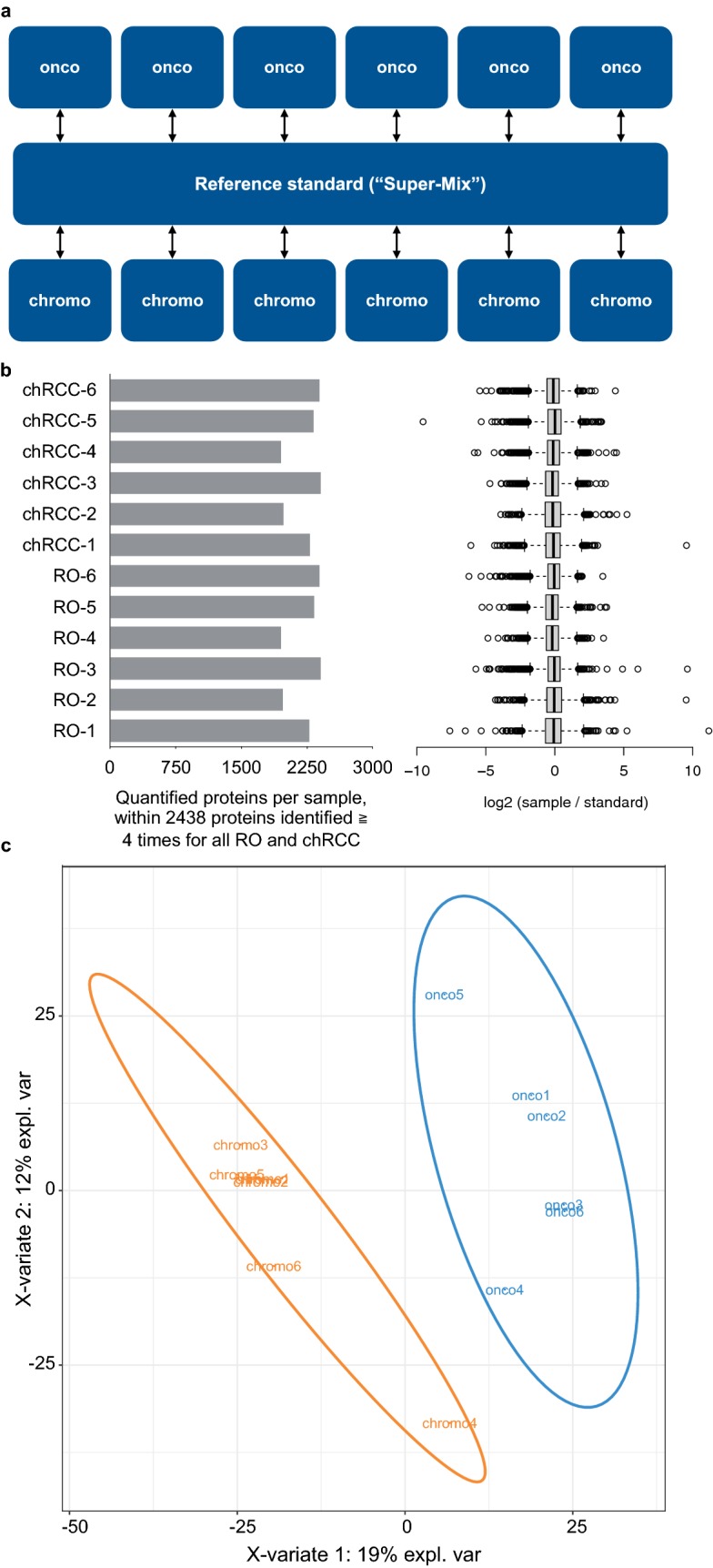
We employed formalin-fixed, paraffin-embedded samples (six RO cases, six chRCC cases) together with isotopic triplex dimethylation and a pooled reference standard to enable cohort-wide quantitative comparison. For lysosomal-associated membrane protein 1 (LAMP1) and integrin alpha-V (ITGAV) we performed corroborative immunohistochemistry (IHC) in an extended cohort of 42 RO cases and 31 chRCC cases.

RESULTS

At 1% false discovery rate, we identified > 3900 proteins, of which > 2400 proteins were consistently quantified in at least four RO and four chRCC cases. The proteomic expression profiling discriminated ROs and chRCCs and highlighted established features such as accumulation of mitochondrial proteins in ROs together with emphasizing the accumulation of endo-lysosomal proteins in chRCCs. In line with the proteomic data, IHC showed enrichment of LAMP1 in chRCC and of ITGAV in RO.

CONCLUSION

We present one of the first differential proteome profiling studies on ROs and chRCCs and highlight differential abundance of LAMP1 and ITGAV in these renal tumors.

摘要

背景

肾嗜酸细胞瘤(RO)是肾脏的良性上皮性肿瘤,而嫌色肾细胞癌(chRCC)是恶性肾肿瘤。后者占肾肿瘤的5 - 7%。RO和chRCC在分子和组织学上表现出明显的相似性,这使得它们的鉴别具有挑战性。我们旨在对RO和早期chRCC进行差异蛋白质组分析,以便更好地了解区分性的蛋白质模式。

方法

我们使用福尔马林固定、石蜡包埋的样本(6例RO病例,6例chRCC病例)以及同位素三重二甲基化和一个混合参考标准,以实现全队列的定量比较。对于溶酶体相关膜蛋白1(LAMP1)和整合素α-V(ITGAV),我们在42例RO病例和31例chRCC病例的扩展队列中进行了验证性免疫组织化学(IHC)。

结果

在1%的错误发现率下,我们鉴定出>3900种蛋白质,其中>2400种蛋白质在至少4例RO和4例chRCC病例中得到一致定量。蛋白质组表达谱区分了RO和chRCC,并突出了已确定的特征,如RO中线粒体蛋白的积累,同时强调了chRCC中内溶酶体蛋白的积累。与蛋白质组数据一致,IHC显示chRCC中LAMP1富集,RO中ITGAV富集。

结论

我们展示了首批关于RO和chRCC的差异蛋白质组分析研究之一,并突出了这些肾肿瘤中LAMP1和ITGAV的差异丰度。

https://cdn.ncbi.nlm.nih.gov/pmc/blobs/3c2e/6074034/61ffb909537d/12014_2018_9200_Fig4_HTML.jpg
https://cdn.ncbi.nlm.nih.gov/pmc/blobs/3c2e/6074034/bbbf23a085b3/12014_2018_9200_Fig1_HTML.jpg
https://cdn.ncbi.nlm.nih.gov/pmc/blobs/3c2e/6074034/ed355a6d438e/12014_2018_9200_Fig3_HTML.jpg
https://cdn.ncbi.nlm.nih.gov/pmc/blobs/3c2e/6074034/61ffb909537d/12014_2018_9200_Fig4_HTML.jpg
https://cdn.ncbi.nlm.nih.gov/pmc/blobs/3c2e/6074034/bbbf23a085b3/12014_2018_9200_Fig1_HTML.jpg
https://cdn.ncbi.nlm.nih.gov/pmc/blobs/3c2e/6074034/ed355a6d438e/12014_2018_9200_Fig3_HTML.jpg
https://cdn.ncbi.nlm.nih.gov/pmc/blobs/3c2e/6074034/61ffb909537d/12014_2018_9200_Fig4_HTML.jpg

相似文献

1
Proteomic distinction of renal oncocytomas and chromophobe renal cell carcinomas.肾嗜酸细胞瘤与嫌色性肾细胞癌的蛋白质组学鉴别
Clin Proteomics. 2018 Aug 3;15:25. doi: 10.1186/s12014-018-9200-6. eCollection 2018.
2
The Utility of ERBB4 and RB1 Immunohistochemistry in Distinguishing Chromophobe Renal Cell Carcinoma From Renal Oncocytoma.ERBB4和RB1免疫组化在鉴别嫌色性肾细胞癌与肾嗜酸细胞瘤中的应用
Int J Surg Pathol. 2020 May;28(3):259-264. doi: 10.1177/1066896919883016. Epub 2019 Oct 22.
3
Use of specific contrast-enhanced CT regions of interest to differentiate renal oncocytomas from small clear cell and chromophobe renal cell carcinomas.使用特定的增强 CT 感兴趣区来区分肾嗜酸细胞瘤与小透明细胞和嫌色细胞肾细胞癌。
Diagn Interv Radiol. 2022 Nov;28(6):555-562. doi: 10.5152/dir.2022.2111504.
4
Sporadic oncocytic tumors with features intermediate between oncocytoma and chromophobe renal cell carcinoma: comprehensive clinicopathological and genomic profiling.散发性具有嗜酸性细胞瘤和嫌色细胞肾细胞癌之间特征的肿瘤:全面的临床病理和基因组分析。
Hum Pathol. 2020 Oct;104:18-29. doi: 10.1016/j.humpath.2020.07.003. Epub 2020 Jul 13.
5
TGF-β1 expression in chromophobe renal cell carcinoma and renal oncocytoma.嗜铬细胞瘤和肾嗜酸细胞瘤中转化生长因子-β1的表达
Eur J Histochem. 2014 Jan 31;58(1):2265. doi: 10.4081/ejh.2014.2265.
6
NKX6-1 Is a Less Sensitive But Specific Biomarker of Chromophobe Renal Cell Carcinoma.NKX6-1是嫌色性肾细胞癌敏感性较低但具有特异性的生物标志物。
Am J Surg Pathol. 2022 Jun 1;46(6):809-815. doi: 10.1097/PAS.0000000000001872. Epub 2022 Mar 8.
7
HNF1β and S100A1 are useful biomarkers for distinguishing renal oncocytoma and chromophobe renal cell carcinoma in FNA and core needle biopsies.在细针穿刺活检(FNA)和粗针穿刺活检中,肝细胞核因子1β(HNF1β)和S100A1是区分肾嗜酸细胞瘤和嫌色性肾细胞癌的有用生物标志物。
Cancer Cytopathol. 2015 May;123(5):298-305. doi: 10.1002/cncy.21530. Epub 2015 Mar 4.
8
Proteomic analysis of chromophobe renal cell carcinoma and benign renal oncocytoma biopsies reveals shared metabolic dysregulation.嫌色性肾细胞癌和良性肾嗜酸细胞瘤活检组织的蛋白质组学分析揭示了共同的代谢失调。
Clin Proteomics. 2023 Nov 28;20(1):54. doi: 10.1186/s12014-023-09443-8.
9
Differential immunohistochemical and molecular profiling of conventional and aggressive components of chromophobe renal cell carcinoma: pitfalls for diagnosis.嫌色细胞肾细胞癌传统成分与侵袭性成分的免疫组化及分子特征差异:诊断陷阱
Hum Pathol. 2022 Jan;119:85-93. doi: 10.1016/j.humpath.2021.11.003. Epub 2021 Nov 17.
10
Chromosome 1 analysis in chromophobe renal cell carcinomas with tissue microarray (TMA)-facilitated fluorescence in situ hybridization (FISH) demonstrates loss of 1p/1 which is also present in renal oncocytomas.利用组织微阵列(TMA)辅助荧光原位杂交(FISH)对嫌色性肾细胞癌进行1号染色体分析,结果显示存在1p/1缺失,这在肾嗜酸细胞瘤中也有出现。
Diagn Mol Pathol. 2008 Sep;17(3):141-4. doi: 10.1097/PDM.0b013e3181577d57.

引用本文的文献

1
Comprehensive proteogenomic characterization of rare kidney tumors.罕见肾脏肿瘤的全面蛋白质基因组学特征分析。
Cell Rep Med. 2024 May 21;5(5):101547. doi: 10.1016/j.xcrm.2024.101547. Epub 2024 May 3.
2
Absolute quantitative proteomics using the total protein approach to identify novel clinical immunohistochemical markers in renal neoplasms.采用全蛋白质法的绝对定量蛋白质组学鉴定肾肿瘤中新型临床免疫组织化学标志物。
BMC Med. 2021 Sep 6;19(1):196. doi: 10.1186/s12916-021-02071-9.
3
Histo-molecular differentiation of renal cancer subtypes by mass spectrometry imaging and rapid proteome profiling of formalin-fixed paraffin-embedded tumor tissue sections.

本文引用的文献

1
Proteomic Characterization of Prostate Cancer to Distinguish Nonmetastasizing and Metastasizing Primary Tumors and Lymph Node Metastases.前列腺癌蛋白质组学特征分析用于鉴别非转移和转移的原发肿瘤及淋巴结转移。
Neoplasia. 2018 Feb;20(2):140-151. doi: 10.1016/j.neo.2017.10.009. Epub 2017 Dec 14.
2
Proteome profiling of clear cell renal cell carcinoma in von Hippel-Lindau patients highlights upregulation of Xaa-Pro aminopeptidase-1, an anti-proliferative and anti-migratory exoprotease.对冯·希佩尔-林道病患者的透明细胞肾细胞癌进行蛋白质组分析,结果显示Xaa-Pro氨肽酶-1上调,这是一种具有抗增殖和抗迁移作用的外切蛋白酶。
Oncotarget. 2017 Oct 19;8(59):100066-100078. doi: 10.18632/oncotarget.21929. eCollection 2017 Nov 21.
3
通过质谱成像和福尔马林固定石蜡包埋肿瘤组织切片的快速蛋白质组分析对肾癌亚型进行组织分子鉴别
Oncotarget. 2020 Nov 3;11(44):3998-4015. doi: 10.18632/oncotarget.27787.
4
Proteomic approaches for characterizing renal cell carcinoma.用于表征肾细胞癌的蛋白质组学方法。
Clin Proteomics. 2020 Jul 29;17:28. doi: 10.1186/s12014-020-09291-w. eCollection 2020.
5
Quantitative Proteomics Using Formalin-fixed, Paraffin-embedded Biopsy Tissues in Inflammatory Disease.使用福尔马林固定、石蜡包埋活检组织进行炎症性疾病的定量蛋白质组学研究
J Proteomics Bioinform. 2019;12(7):104-112. doi: 10.35248/0974-276X.12.19.503. Epub 2019 Oct 3.
6
Identification of as a novel key gene in chromophobe renal cell carcinoma through bioinformatics analysis.通过生物信息学分析鉴定某基因作为嫌色性肾细胞癌中的一个新的关键基因。 (注:原文中Identification of后面缺少具体基因名称,这里翻译为“某基因”,实际翻译时应根据准确的原文内容进行完整准确翻译)
Oncol Lett. 2019 Aug;18(2):1767-1774. doi: 10.3892/ol.2019.10476. Epub 2019 Jun 14.
A pathology atlas of the human cancer transcriptome.
人类癌症转录组病理学图谱。
Science. 2017 Aug 18;357(6352). doi: 10.1126/science.aan2507.
4
Can we predict protein from mRNA levels?我们能否根据mRNA水平预测蛋白质?
Nature. 2017 Jul 26;547(7664):E19-E20. doi: 10.1038/nature22293.
5
On the Dependency of Cellular Protein Levels on mRNA Abundance.细胞蛋白质水平对mRNA丰度的依赖性
Cell. 2016 Apr 21;165(3):535-50. doi: 10.1016/j.cell.2016.03.014.
6
A systematic review and meta-analysis of immunohistochemical biomarkers that differentiate chromophobe renal cell carcinoma from renal oncocytoma.鉴别嫌色性肾细胞癌与肾嗜酸细胞瘤的免疫组化生物标志物的系统评价和荟萃分析
J Clin Pathol. 2016 Aug;69(8):661-71. doi: 10.1136/jclinpath-2015-203585. Epub 2016 Mar 7.
7
Multilevel Genomics-Based Taxonomy of Renal Cell Carcinoma.基于多组学的肾细胞癌分类法
Cell Rep. 2016 Mar 15;14(10):2476-89. doi: 10.1016/j.celrep.2016.02.024. Epub 2016 Mar 3.
8
The Genomic Landscape of Renal Oncocytoma Identifies a Metabolic Barrier to Tumorigenesis.肾嗜酸细胞瘤的基因组图谱揭示了肿瘤发生的代谢障碍。
Cell Rep. 2015 Dec 1;13(9):1895-908. doi: 10.1016/j.celrep.2015.10.059. Epub 2015 Nov 19.
9
2016 update of the PRIDE database and its related tools.PRIDE数据库及其相关工具的2016年更新。
Nucleic Acids Res. 2016 Jan 4;44(D1):D447-56. doi: 10.1093/nar/gkv1145. Epub 2015 Nov 2.
10
Impact of cathepsin B on the interstitial fluid proteome of murine breast cancers.组织蛋白酶B对小鼠乳腺癌间质液蛋白质组的影响。
Biochimie. 2016 Mar;122:88-98. doi: 10.1016/j.biochi.2015.10.009. Epub 2015 Oct 8.