Department of Biomedical Engineering, Northwestern University, Evanston, IL, 60208, USA.
Center for Endoscopic Research and Therapeutics, University of Chicago Medicine, Chicago, IL, 60637, USA.
BMC Cancer. 2018 Aug 13;18(1):814. doi: 10.1186/s12885-018-4709-7.
The present study aimed to investigate the role of blood supply in early tumorigenesis in colorectal cancer. We leveraged the renin angiotensin system (RAS) to alter colonic blood supply and determine the effect on tumor initiation and progression.
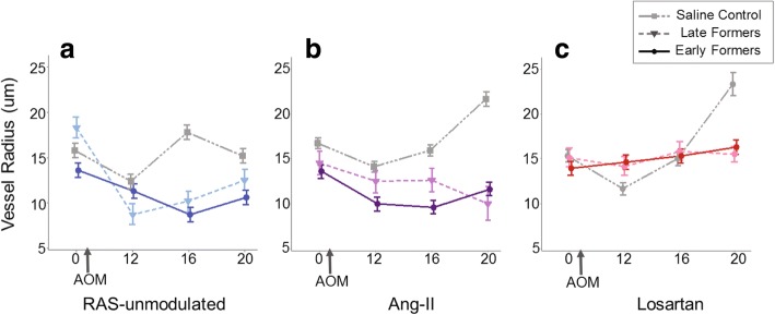
To test the effect of blood supply on tumorigenesis, 53 male A/J mice were randomly assigned to one of three RAS modulation groups and one of two AOM treatments. The RAS modulation groups were I) water (RAS-unmodulated) as a control group, II) angiotensin-II and III) the angiotensin receptor blocker, Losartan. The mice in each group were then randomly split into either the saline control condition or the AOM-treated condition in which tumors were induced with a standard protocol of serial azoxymethane (AOM) injections. To monitor microvascular changes in the rectal mucosa during the study, we used confocal laser endomicroscopy (CLE) with FITC-Dextran for in-vivo imaging of vessels and polarization-gated spectroscopy (PGS) to quantify rectal hemoglobin concentration ([Hb]) and blood vessel radius (BVR).
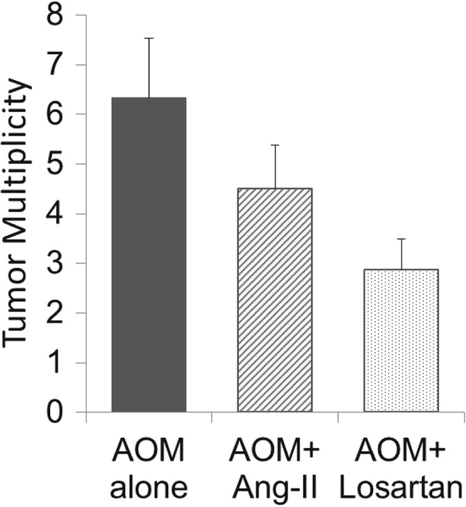
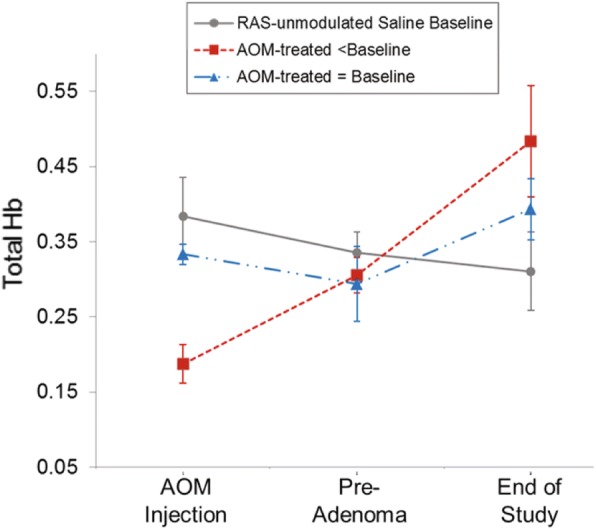
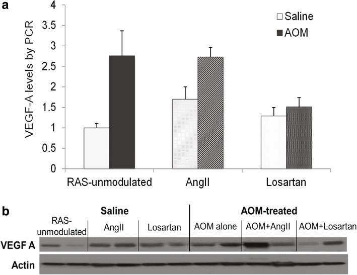
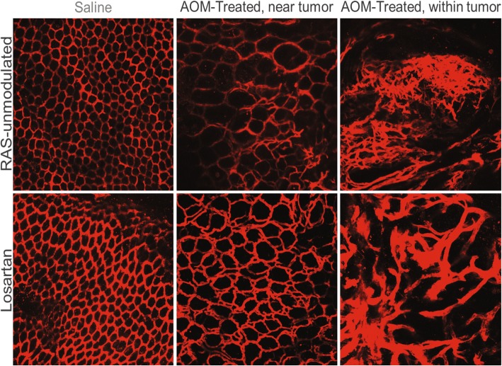
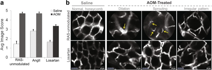
At 12 weeks post-AOM injections and before tumor formation, CLE images revealed many traditional hallmarks of angiogenesis including vessel dilation, loss of co-planarity, irregularity, and vessel sprouting in the pericryptal capillaries of the rectal mucosa in AOM-Water tumor bearing mice. PGS measurements at the same time-point showed increased rectal [Hb] and decreased BVR. At later time points, CLE images showed pronounced angiogenic features including irregular networks throughout the colon. Notably, the AOM-Losartan mice had significantly lower tumor multiplicity and did not exhibit the same angiogenic features observed with CLE, or the increase in [Hb] or decrease in BVR measured with PGS. The AOM-AngII mice did not have any significant trends.
In-vivo PGS measurements of rectal colonic blood supply as well as CLE imaging revealed angiogenic disruptions to the capillary network prior to tumor formation. Losartan demonstrated an effective way to mitigate the changes to blood supply during tumorigenesis and reduce tumor multiplicity. These effects can be used in future studies to understand the early vessel changes observed.
本研究旨在探讨结直肠癌早期肿瘤发生过程中血液供应的作用。我们利用肾素-血管紧张素系统(RAS)改变结肠血液供应,并确定其对肿瘤起始和进展的影响。
为了研究血液供应对肿瘤发生的影响,将 53 只雄性 A/J 小鼠随机分为三个 RAS 调节组和两个 AOM 处理组之一。RAS 调节组为 I)水(RAS 未调节)作为对照组,II)血管紧张素-II 和 III)血管紧张素受体阻滞剂,氯沙坦。然后,每组小鼠随机分为生理盐水对照组或 AOM 处理组,采用标准的偶氮甲烷(AOM)注射系列诱导肿瘤。为了在研究过程中监测直肠黏膜微血管变化,我们使用带有 FITC-右旋糖酐的共聚焦激光内窥显微镜(CLE)进行血管的体内成像和偏振门控光谱(PGS)来定量直肠血红蛋白浓度([Hb])和血管半径(BVR)。
在 AOM 注射后 12 周,即在肿瘤形成之前,CLE 图像显示,在 AOM-水荷瘤小鼠的直肠黏膜血管周围毛细血管中,许多传统的血管生成标志物包括血管扩张、共面性丧失、不规则性和血管发芽。在同一时间点的 PGS 测量显示直肠[Hb]增加和 BVR 降低。在稍后的时间点,CLE 图像显示出明显的血管生成特征,包括整个结肠不规则的网络。值得注意的是,AOM-氯沙坦组的肿瘤多发性显著降低,并且没有表现出 CLE 观察到的相同的血管生成特征,或 PGS 测量的[Hb]增加或 BVR 降低。AOM-血管紧张素 II 组没有任何明显的趋势。
直肠结肠血液供应的体内 PGS 测量以及 CLE 成像在肿瘤形成前揭示了毛细血管网络的血管生成破坏。氯沙坦在肿瘤发生过程中为减轻血液供应的变化并降低肿瘤多发性提供了一种有效的方法。这些作用可用于未来的研究中,以了解观察到的早期血管变化。