Department of Physiology, University of Louisville, 505 South Hancock Street, CTRB, Room 322, Louisville, 40202, KY, USA.
James Graham Brown Cancer Center, University of Louisville, Louisville, 40202, KY, USA.
J Ovarian Res. 2018 Aug 18;11(1):69. doi: 10.1186/s13048-018-0439-3.
Ovarian cancer is a complicated malady associated with cancer stem cells (CSCs) contributing to 238,700 estimated new cases and 151,900 deaths per year, worldwide. CSCs comprise a tiny fraction of tumor-bulk responsible for cancer recurrence and eventual mortality. CSCs or tumor initiating cells are responsible for self-renewal, differentiation and proliferative potential, tumor initiation capability, its progression, drug resistance and metastatic spread. Although several biomarkers are implicated in these processes, their distribution within the ovary and association with single cell type has neither been established nor demonstrated across ovarian tumor developmental stages. Therefore, precise identification, thorough characterization and effective targeted destruction of dormant and highly proliferating potent CSC populations is an immediate need.
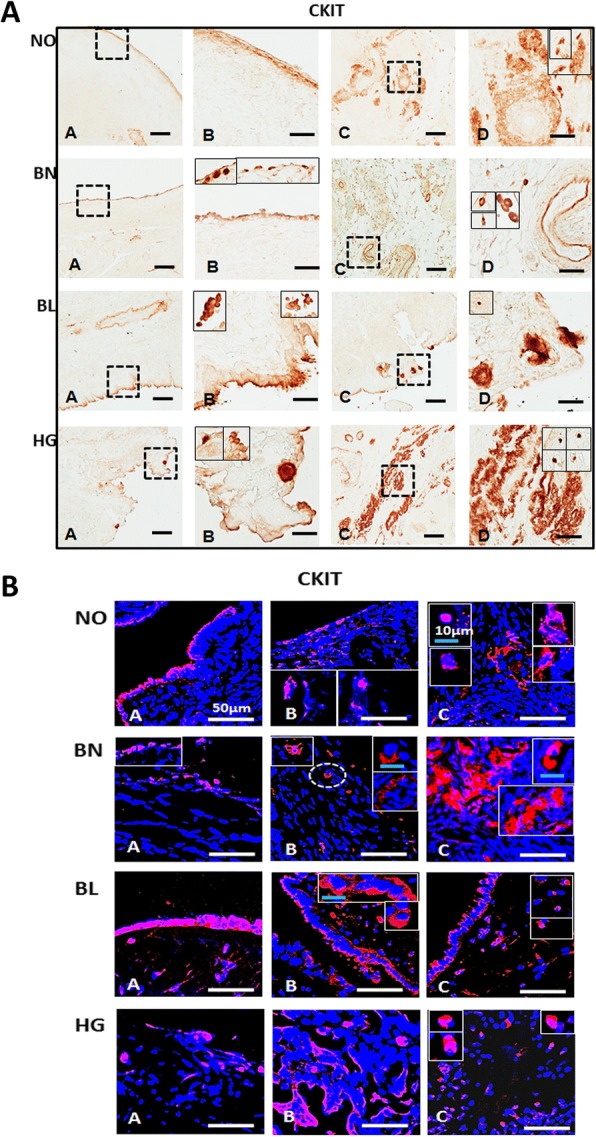
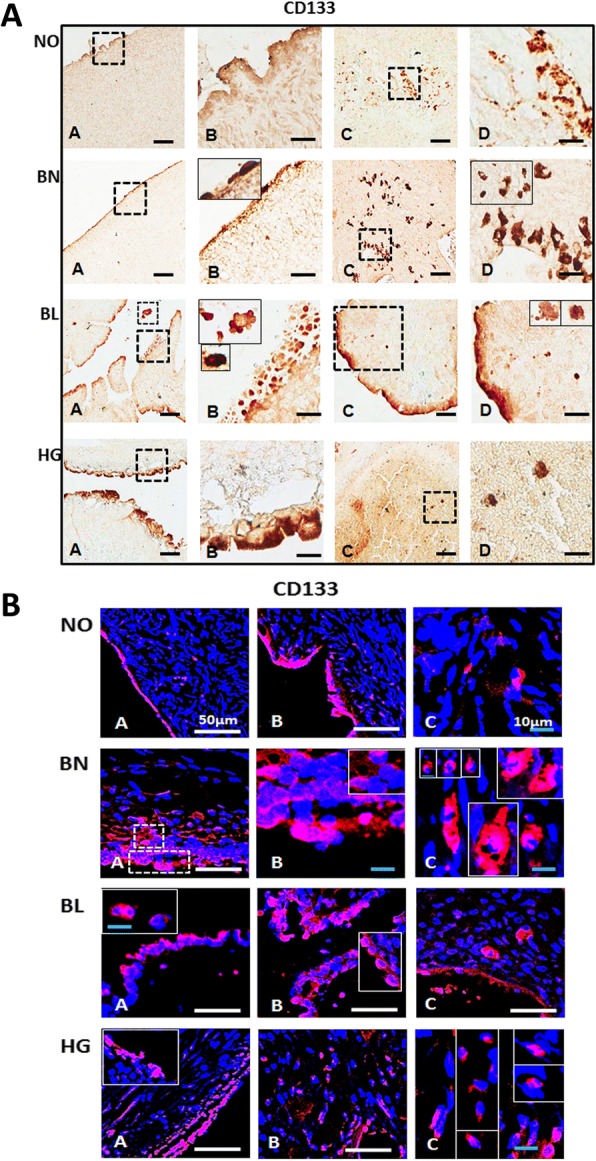
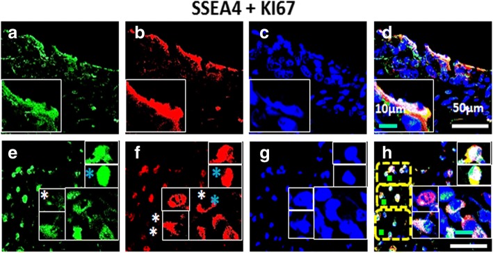
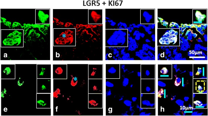
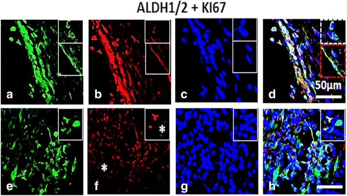
In view of this, distribution of various CSC (ALDH1/2, C-KIT, CD133, CD24 and CD44) and cell proliferation (KI67) specific markers in the ovarian surface epithelium (OSE) and cortex regions in normal ovary, and benign, borderline and high grade metastatic ovarian tumors by immuno-histochemistry and confocal microscopy was studied. We further confirmed their expression by RT-PCR analysis. Co-expression analysis of stem cell (OCT4, SSEA4) and CSC (ALDH1/2, CD44 and LGR5) markers with proliferation marker (KI67) in HG tumors revealed dual positive proliferating stem and CSCs, few non-proliferating stem/CSC (SSEA4/KI67 and ALDH1/2/KI67) and only KI67 cells in cortex, signifying dynamic populations and interesting cellular hierarchy in cortex region. Smaller spherical (≤ 5 μm) and larger spindle/elliptical shaped (~ 10 μm) cell populations with high nucleo-cytoplasmic ratio were detected across all samples (including normal ovaries) but with variable distribution and characteristic stage-wise marker expression across different tumor stages.
Diverse stem and CSC populations expressing characteristic markers revealing distinct phenotypes (spherical ≤5 μm and spindle/elliptical ~ 10 μm) were distributed within different tumor stages studied signifying dynamic and probable functional hierarchy within these cell types. Involvement of extra-ovarian sites of origin of stem and CSCs requires rigorous evaluation. Quantitative analysis of potent CSC populations, their mechanisms and pathways for self-renewal, chemo-resistance, metastatic spread etc. with respect to various markers studied, will provide better insights and targets for developing effective therapeutics to prevent metastasis and eventually help improve patient mortality.
卵巢癌是一种复杂的疾病,与癌症干细胞(CSCs)有关,全球每年估计有 238700 例新发病例和 151900 例死亡。CSCs 仅占肿瘤实体的一小部分,是导致癌症复发和最终死亡的原因。CSCs 或肿瘤起始细胞负责自我更新、分化和增殖潜能、肿瘤起始能力、进展、耐药性和转移扩散。尽管有几种生物标志物与这些过程有关,但它们在卵巢内的分布及其与单个细胞类型的关联尚未在卵巢肿瘤发育的各个阶段得到证实。因此,精确识别、彻底表征和有效靶向休眠和高增殖的潜在 CSC 群体是当务之急。
有鉴于此,我们通过免疫组织化学和共聚焦显微镜研究了各种 CSC(ALDH1/2、C-KIT、CD133、CD24 和 CD44)和细胞增殖(KI67)特异性标志物在正常卵巢的卵巢表面上皮(OSE)和皮质区域,以及良性、交界性和高级转移性卵巢肿瘤中的分布。我们还通过 RT-PCR 分析证实了它们的表达。在 HG 肿瘤中,与增殖标志物(KI67)共表达的干细胞(OCT4、SSEA4)和 CSC(ALDH1/2、CD44 和 LGR5)标志物的分析表明,有双阳性增殖的干细胞和 CSC、少数非增殖的干细胞/CSC(SSEA4/KI67 和 ALDH1/2/KI67)和仅在皮质区的 KI67 细胞,这表明在皮质区存在动态群体和有趣的细胞层次结构。在所有样本(包括正常卵巢)中都检测到较小的球形(≤5μm)和较大的纺锤形/椭圆形(~10μm)细胞群体,具有较高的核质比,但在不同肿瘤阶段的分布和特征性阶段性标志物表达存在差异。
在研究的不同肿瘤阶段内,分布着表达特征性标志物的不同的干细胞和 CSC 群体,这些标志物揭示了不同的表型(≤5μm 的球形和~10μm 的纺锤形/椭圆形),这表明这些细胞类型中存在动态和可能的功能层次结构。需要严格评估卵巢外起源的干细胞和 CSC 部位的参与。对具有不同标志物的潜在 CSC 群体的定量分析、它们的自我更新、化疗耐药性、转移扩散等机制和途径,将为开发有效的治疗方法以预防转移并最终帮助提高患者生存率提供更好的见解和目标。