• 文献检索
  • 文档翻译
  • 深度研究
  • 学术资讯
  • Suppr Zotero 插件Zotero 插件
  • 邀请有礼
  • 套餐&价格
  • 历史记录
应用&插件
Suppr Zotero 插件Zotero 插件浏览器插件Mac 客户端Windows 客户端微信小程序
定价
高级版会员购买积分包购买API积分包
服务
文献检索文档翻译深度研究API 文档MCP 服务
关于我们
关于 Suppr公司介绍联系我们用户协议隐私条款
关注我们

Suppr 超能文献

核心技术专利:CN118964589B侵权必究
粤ICP备2023148730 号-1Suppr @ 2026

文献检索

告别复杂PubMed语法,用中文像聊天一样搜索,搜遍4000万医学文献。AI智能推荐,让科研检索更轻松。

立即免费搜索

文件翻译

保留排版,准确专业,支持PDF/Word/PPT等文件格式,支持 12+语言互译。

免费翻译文档

深度研究

AI帮你快速写综述,25分钟生成高质量综述,智能提取关键信息,辅助科研写作。

立即免费体验

Central retinal vein occlusion revealing celiac disease: The first report of two cases from India.

作者信息

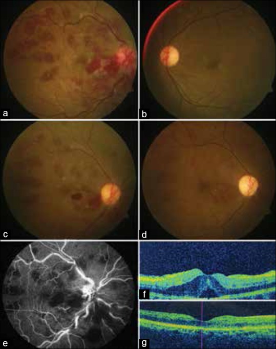
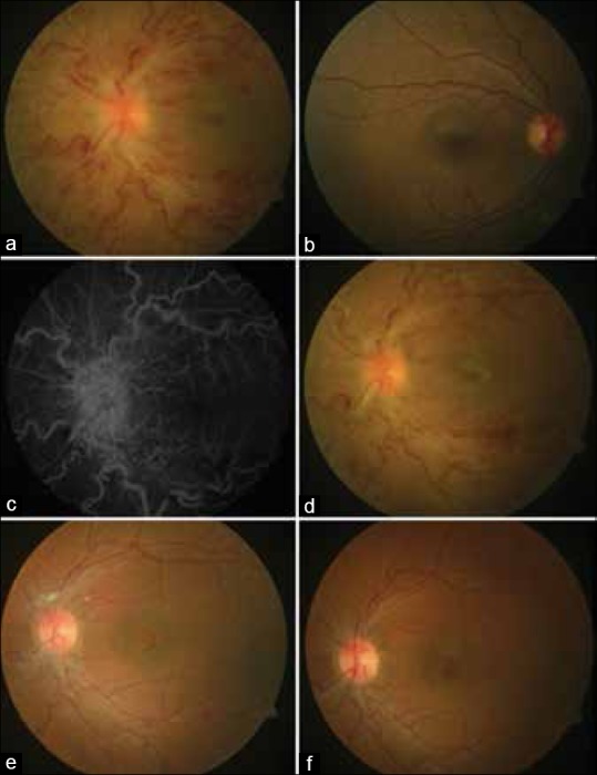
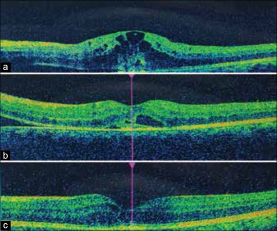
Malhi Ravinder Kaur, Dhami Abhinav, Malhi Nirmaljeet Singh, Soni Amit, Dhami Gobinder Singh

机构信息

Department of Vitreo-Retina, Dhami Eye Care Hospital, Ludhiana, Punjab, India.

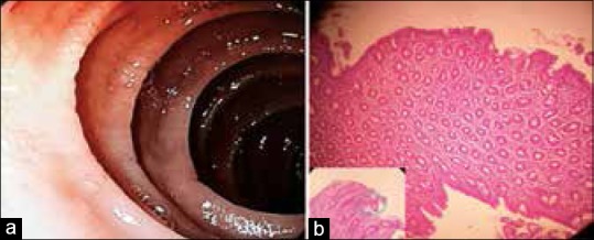
Department of Gastroenterology and Liver Diseases, SPS Hospital, Ludhiana, Punjab, India.

出版信息

Indian J Ophthalmol. 2018 Sep;66(9):1315-1317. doi: 10.4103/ijo.IJO_351_18.

DOI:10.4103/ijo.IJO_351_18
PMID:30127151
原文链接:https://pmc.ncbi.nlm.nih.gov/articles/PMC6113809/
Abstract
摘要
https://cdn.ncbi.nlm.nih.gov/pmc/blobs/7277/6113809/292c5cf3f2e7/IJO-66-1315-g005.jpg
https://cdn.ncbi.nlm.nih.gov/pmc/blobs/7277/6113809/97833396950b/IJO-66-1315-g001.jpg
https://cdn.ncbi.nlm.nih.gov/pmc/blobs/7277/6113809/4d1f82b8117a/IJO-66-1315-g002.jpg
https://cdn.ncbi.nlm.nih.gov/pmc/blobs/7277/6113809/f5be8ce5ae58/IJO-66-1315-g003.jpg
https://cdn.ncbi.nlm.nih.gov/pmc/blobs/7277/6113809/292c5cf3f2e7/IJO-66-1315-g005.jpg

相似文献

1
Central retinal vein occlusion revealing celiac disease: The first report of two cases from India.中央视网膜静脉阻塞揭示乳糜泻:来自印度的两例首例报告。
Indian J Ophthalmol. 2018 Sep;66(9):1315-1317. doi: 10.4103/ijo.IJO_351_18.
2
Optical coherence tomography angiography wide-field montage in branch retinal vein occlusion before and after anti-vascular endothelial-derived growth factor injection.抗血管内皮生长因子注射前后视网膜分支静脉阻塞的光学相干断层扫描血管造影宽视野拼接成像
Int Ophthalmol. 2018 Jun;38(3):1305-1307. doi: 10.1007/s10792-017-0568-5. Epub 2017 May 24.
3
Swept-Source OCT Angiographic Imaging of a Central Retinal Vein Occlusion During Pregnancy.妊娠期视网膜中央静脉阻塞的扫频源光学相干断层扫描血管造影成像
Ophthalmic Surg Lasers Imaging Retina. 2018 Mar 1;49(3):206-208. doi: 10.3928/23258160-20180221-09.
4
Bilateral combined central retinal artery and vein occlusion in a 3-year-old child with nephrotic syndrome.3 岁肾病综合征患儿合并双侧中央视网膜动脉和静脉阻塞。
Indian J Ophthalmol. 2018 Oct;66(10):1498-1501. doi: 10.4103/ijo.IJO_1332_17.
5
QUALITATIVE AND QUANTITATIVE FOLLOW-UP USING OPTICAL COHERENCE TOMOGRAPHY ANGIOGRAPHY OF RETINAL VEIN OCCLUSION TREATED WITH ANTI-VEGF: Optical Coherence Tomography Angiography Follow-up of Retinal Vein Occlusion.使用抗血管内皮生长因子治疗的视网膜静脉阻塞的光学相干断层扫描血管造影的定性和定量随访:视网膜静脉阻塞的光学相干断层扫描血管造影随访
Retina. 2017 Jun;37(6):1176-1184. doi: 10.1097/IAE.0000000000001334.
6
Bilateral simultaneous retinal vein occlusion.
Indian J Ophthalmol. 1999 Dec;47(4):248-50.
7
Ischemic central retinal vein occlusion in a 14-year-old female.一名14岁女性的缺血性视网膜中央静脉阻塞。
Can J Ophthalmol. 2017 Apr;52(2):e64-e66. doi: 10.1016/j.jcjo.2016.09.012. Epub 2017 Jan 27.
8
Central Retinal Vein Occlusion with Chorioretinal Folds Secondary to Active Thyroid Eye Disease.继发于活动性甲状腺眼病的伴有脉络膜视网膜皱褶的视网膜中央静脉阻塞
Ophthalmology. 2018 Oct;125(10):1645. doi: 10.1016/j.ophtha.2018.07.001.
9
Retinal vessel tortuosity associated with central retinal vein occlusion: an optical coherence tomography study.视网膜血管迂曲与视网膜中央静脉阻塞的关系:一项光学相干断层扫描研究。
Invest Ophthalmol Vis Sci. 2014 Jan 7;55(1):134-41. doi: 10.1167/iovs.13-13201.
10
Retinal Arteriovenous Malformation Occlusion and Optic Nerve Drusen: Casuality or Causality?视网膜动静脉畸形闭塞与视神经玻璃疣:偶然还是必然?
Ophthalmic Surg Lasers Imaging Retina. 2020 Jul 1;51(7):418-419. doi: 10.3928/23258160-20200702-08.

引用本文的文献

1
Combined Central Retinal Artery Occlusion (CRAO) and Central Retinal Vein Occlusion (CRVO) in a Celiac Disease Patient: A Case Report.一名腹腔疾病患者合并视网膜中央动脉阻塞(CRAO)和视网膜中央静脉阻塞(CRVO):病例报告
Cureus. 2024 Jan 3;16(1):e51567. doi: 10.7759/cureus.51567. eCollection 2024 Jan.
2
Choroidal structural evaluation in celiac disease.腹腔疾病的脉络膜结构评估。
Sci Rep. 2021 Aug 12;11(1):16398. doi: 10.1038/s41598-021-95812-y.
3
Ocular manifestations in celiac disease: an overview.腹腔疾病的眼部表现:概述。

本文引用的文献

1
[An atypical presentation of celiac disease: central retinal vein occlusion].[乳糜泻的非典型表现:视网膜中央静脉阻塞]
Pan Afr Med J. 2015 Nov 25;22:300. doi: 10.11604/pamj.2015.22.300.8196. eCollection 2015.
2
Ophthalmologic manifestations of celiac disease.乳糜泻的眼科表现。
Int J Ophthalmol. 2016 Jan 18;9(1):159-62. doi: 10.18240/ijo.2016.01.26. eCollection 2016.
3
Celiac disease in India.印度的乳糜泻
Int Ophthalmol. 2020 Apr;40(4):1049-1054. doi: 10.1007/s10792-019-01254-x. Epub 2020 Jan 8.
4
Celiac Disease: Extraintestinal Manifestations and Associated Conditions.乳糜泻:肠外表现和相关疾病。
J Clin Gastroenterol. 2020 Jan;54(1):8-21. doi: 10.1097/MCG.0000000000001267.
Indian J Gastroenterol. 2007 Sep-Oct;26(5):230-7.
4
Nonischemic central retinal vein occlusion associated with celiac disease.与腹腔疾病相关的非缺血性视网膜中央静脉阻塞
Mayo Clin Proc. 2005 Feb;80(2):157. doi: 10.4065/80.2.157.