Myocardial Function Section, Imperial Centre for Translational and Experimental Medicine, Imperial College London, London, UK.
Imperial Centre for Cardiac Engineering, Imperial College London, London, UK.
J Cardiovasc Electrophysiol. 2018 Dec;29(12):1624-1634. doi: 10.1111/jce.13723. Epub 2018 Oct 5.
The ganglionated plexuses (GPs) of the intrinsic cardiac autonomic system are implicated in arrhythmogenesis. GP localization by stimulation of the epicardial fat pads to produce atrioventricular dissociating (AVD) effects is well described. We determined the anatomical distribution of the left atrial GPs that influence atrioventricular (AV) dissociation.
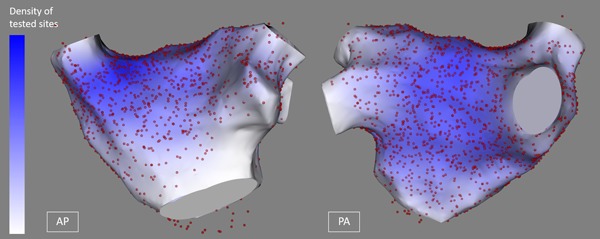
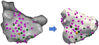
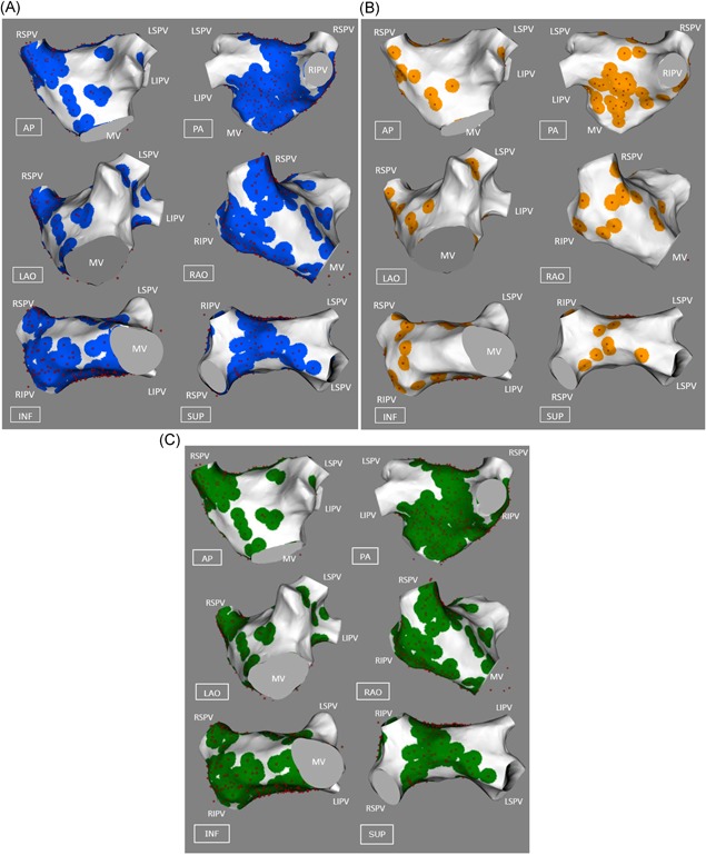
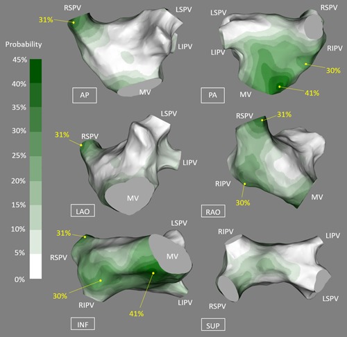
High frequency stimulation was delivered through a Smart-Touch catheter in the left atrium of patients undergoing atrial fibrillation (AF) ablation. Three dimensional locations of points tested throughout the entire chamber were recorded on the CARTO™ system. Impact on the AV conduction was categorized as ventricular asystole, bradycardia, or no effect. CARTO maps were exported, registered, and transformed onto a reference left atrial geometry using a custom software, enabling data from multiple patients to be overlaid. In 28 patients, 2108 locations were tested and 283 sites (13%) demonstrated (AVD-GP) effects. There were 10 AVD-GPs (interquartile range, 11.5) per patient. Eighty percent (226) produced asystole and 20% (57) showed bradycardia. The distribution of the two groups was very similar. Highest probability of AVD-GPs (>20%) was identified in: inferoseptal portion (41%) and right inferior pulmonary vein base (30%) of the posterior wall, right superior pulmonary vein antrum (31%).
It is feasible to map the entire left atrium for AVD-GPs before AF ablation. Aggregated data from multiple patients, producing a distribution probability atlas of AVD-GPs, identified three regions with a higher likelihood for finding AVD-GPs and these matched the histological descriptions. This approach could be used to better characterize the autonomic network.
内在心脏自主神经系统的神经节丛(GP)与心律失常的发生有关。通过刺激心外膜脂肪垫来产生房室分离(AVD)作用,已经很好地描述了 GP 的定位。我们确定了影响房室(AV)分离的左房 GP 的解剖分布。
在接受心房颤动(AF)消融的患者的左心房中,通过 Smart-Touch 导管进行高频刺激。在 CARTO™系统上记录整个房间内测试点的三维位置。将对 AV 传导的影响分为心室停搏、心动过缓或无影响。将 CARTO 图谱导出、注册并转换到参考左心房几何形状上,使用自定义软件,从而可以将多个患者的数据叠加。在 28 名患者中,测试了 2108 个部位,283 个部位(13%)显示出(AVD-GP)作用。每位患者有 10 个 AVD-GP(四分位间距,11.5)。80%(226)产生停搏,20%(57)表现为心动过缓。两组的分布非常相似。在后壁的后下间隔部分(41%)和右下肺静脉基底(30%)以及右上肺静脉窦(31%),发现 AVD-GP 的概率最高(>20%)。
在进行 AF 消融之前,对整个左心房进行 AVD-GP 映射是可行的。从多个患者聚合数据,产生 AVD-GP 的分布概率图谱,确定了三个发现 AVD-GP 的可能性更高的区域,这些区域与组织学描述相匹配。这种方法可用于更好地描述自主神经系统网络。