Sims-Williams Helen J, Sims-Williams Hugh P, Mbabazi Kabachelor Edith, Warf Benjamin C
Sheffield Kidney Institute, Sheffield Teaching Hospitals NHS Foundation Trust, Sheffield, UK.
Department of Neurosurgery, Sheffield Teaching Hospitals NHS Foundation Trust, Sheffield, UK.
Int J Nephrol. 2018 Aug 7;2018:6278616. doi: 10.1155/2018/6278616. eCollection 2018.
To describe the extent of renal disease in Ugandan children surviving at least ten years after spina bifida repair and to investigate risk factors for renal deterioration in this cohort.
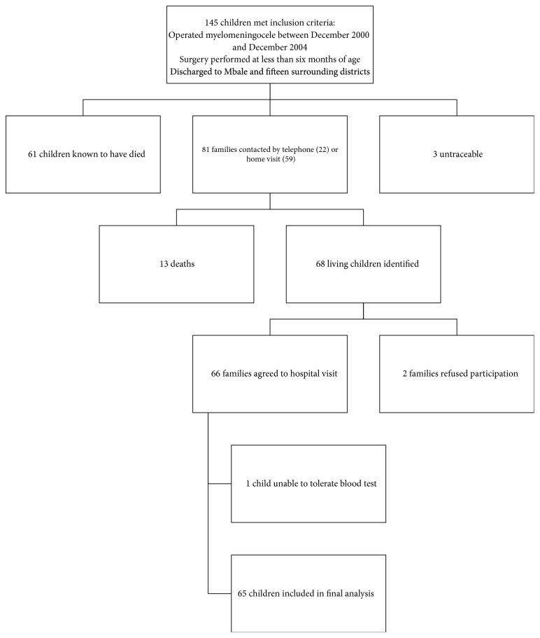
Children who had undergone spina bifida repair at CURE Children's Hospital of Uganda between 2000 and 2004 were invited to attend interview, physical examination, renal tract ultrasound, and a blood test (creatinine). Medical records were retrospectively reviewed. The following were considered evidence of renal damage: elevated creatinine, hypertension, and ultrasound findings of hydronephrosis, scarring, and discrepancy in renal size >1cm. Female sex, previous UTI, neurological level, mobility, detrusor leak point pressure, and adherence with clean intermittent catheterisation (CIC) were investigated for association with evidence of renal damage.
65 of 68 children aged 10-14 completed the assessment. The majority (83%) reported incontinence. 17 children (26%) were performing CIC. One child had elevated creatinine. 25 children (38%) were hypertensive. There was a high prevalence of ultrasound abnormalities: hydronephrosis in 10 children (15%), scarring in 42 (64%), and >1cm size discrepancy in 28 (43%). No children with lesions at S1 or below had hydronephrosis (p = 0.025), but this group had comparable prevalence of renal size discrepancy, scarring, and hypertension to those children with higher lesions.
Incontinence, ultrasound abnormalities, and hypertension are highly prevalent in a cohort of Ugandan children with spina bifida, including those with low neurological lesions. These findings support the early and universal initiation of CIC with anticholinergic therapy in a low-income setting.
描述乌干达脊柱裂修复术后存活至少十年的儿童肾病的严重程度,并调查该队列中肾脏恶化的危险因素。
邀请2000年至2004年间在乌干达CURE儿童医院接受脊柱裂修复术的儿童参加访谈、体格检查、肾超声检查和血液检测(肌酐)。对医疗记录进行回顾性审查。以下情况被视为肾脏损害的证据:肌酐升高、高血压以及肾积水、瘢痕形成和肾大小差异>1cm的超声检查结果。研究女性性别、既往泌尿道感染、神经学水平、活动能力、逼尿肌漏点压力以及清洁间歇性导尿(CIC)依从性与肾脏损害证据之间的关联。
68名10至14岁儿童中的65名完成了评估。大多数(83%)报告有尿失禁。17名儿童(26%)进行CIC。1名儿童肌酐升高。25名儿童(38%)患有高血压。超声异常的患病率很高:10名儿童(15%)有肾积水,42名(64%)有瘢痕形成,28名(43%)肾大小差异>1cm。S1或以下水平有病变的儿童无肾积水(p = 0.025),但该组肾大小差异、瘢痕形成和高血压的患病率与病变较高的儿童相当。
在乌干达脊柱裂儿童队列中,包括神经学病变较轻的儿童,尿失禁、超声异常和高血压非常普遍。这些发现支持在低收入环境中早期普遍开始使用抗胆碱能疗法进行CIC。