Castaneda Katrina A, Hudson Caleb C, Beale Brian S
MedVet Medical and Cancer Centers, 300 E. Wilson Bridge Rd, Worthington, OH, 43085, USA.
Gulf Coast Veterinary Specialists, 1030 Wirt Rd, Houston, TX, 77055, USA.
BMC Vet Res. 2018 Sep 3;14(1):270. doi: 10.1186/s12917-018-1599-5.
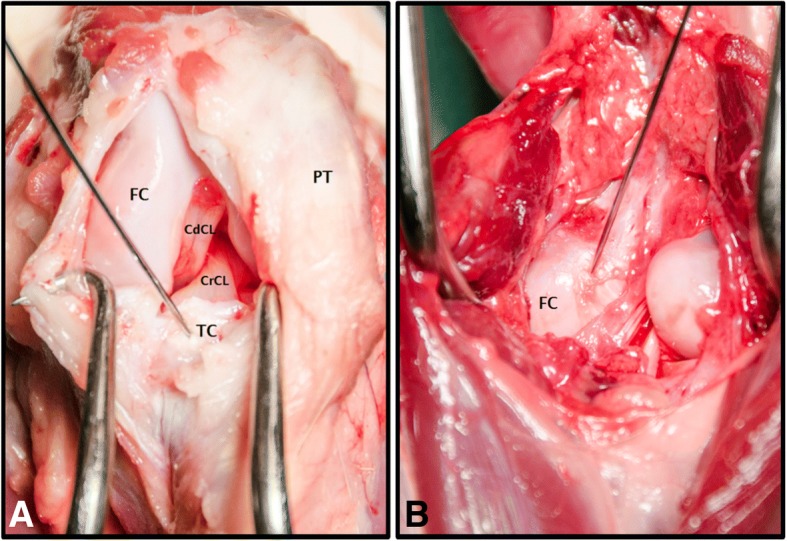
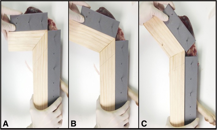
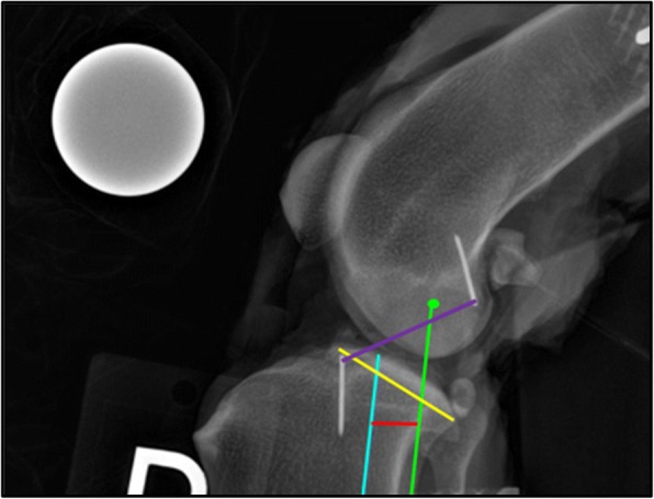
The presence of cranial tibial subluxation can aid in the detection of joint instability as a result of CrCL injury. Detection of cranial tibial subluxation has been described using the tibial compression test (TCT) and cranial drawer test (CDT); however, diagnosis of CrCL insufficiency by assessing cranial subluxation motion of the tibia is subjective and difficult to quantify accurately. The aim of this study was to investigate a measurement technique to assess the degree of cranial tibial displacement relative to the femoral condyles on mediolateral projection stifle radiographs at varying degrees of stifle flexion (90°, 110°, and 135°) in CrCL intact, partially, and completely transected conditions. Radiographic measurements included: CrCL length and intercondylar distance (ICD), defined as the distance between the tibial mechanical axis (TMA) and the femoral condylar axis (FCA). The influence of CrCL status, stifle flexion angle, and measurement type on measured distance was evaluated. The relationship between CrCL length and ICD measurement was also assessed.
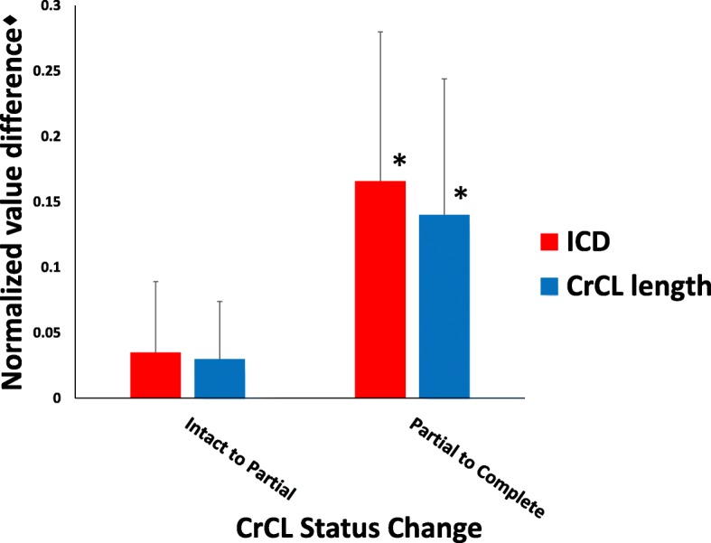
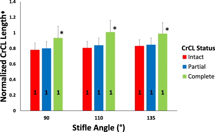
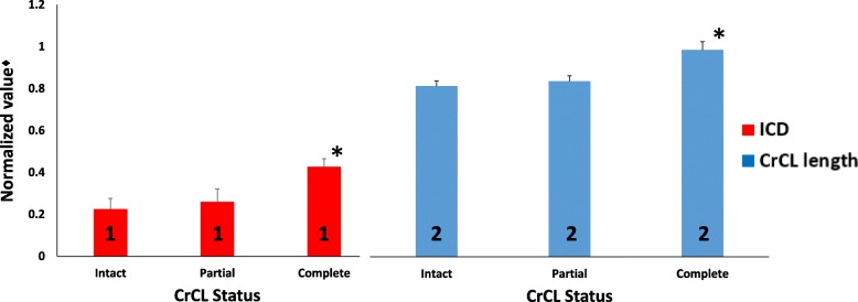
Complete transection of the CrCL resulted in significant cranial tibial displacement. Stifle flexion angle affected ICD, but not CrCL length. Normalized measured CrCL length and ICD were significantly different; however, no differences existed between the change in distance detected by CrCL length and ICD measurements as CrCL transection status changed. Correlation coefficients detected a significant positive correlation between measured CrCL and ICD.
The ICD measurement technique was able to quantify tibial displacement at various stifle flexion angles in the intact and completely transected CrCL conditions. The ICD measurement was more affected by stifle flexion angle than was the CrCL length.
胫骨髁前半脱位的存在有助于检测由前交叉韧带(CrCL)损伤导致的关节不稳定。已通过胫骨挤压试验(TCT)和髁前抽屉试验(CDT)来描述胫骨髁前半脱位的检测;然而,通过评估胫骨髁前半脱位运动来诊断CrCL功能不全具有主观性,且难以准确量化。本研究的目的是探讨一种测量技术,以评估在不同程度的 stifle 屈曲(90°、110°和135°)时,在CrCL完整、部分和完全横断的情况下,在内外侧位 stifle 射线照片上胫骨相对于股骨髁的髁前移位程度。射线照片测量包括:CrCL长度和髁间距离(ICD),定义为胫骨机械轴(TMA)与股骨髁轴(FCA)之间的距离。评估了CrCL状态、stifle屈曲角度和测量类型对测量距离的影响。还评估了CrCL长度与ICD测量之间的关系。
CrCL完全横断导致明显的胫骨髁前移位。stifle屈曲角度影响ICD,但不影响CrCL长度。标准化测量的CrCL长度和ICD有显著差异;然而,随着CrCL横断状态的改变,CrCL长度和ICD测量所检测到的距离变化之间没有差异。相关系数检测到测量的CrCL与ICD之间存在显著正相关。
ICD测量技术能够量化在CrCL完整和完全横断情况下不同stifle屈曲角度的胫骨移位。ICD测量比CrCL长度更容易受到stifle屈曲角度的影响。