From the Department of Structural and Molecular Biology, Darwin Building, University College London, Gower Street, London WC1E 6BT, United Kingdom.
From the Department of Structural and Molecular Biology, Darwin Building, University College London, Gower Street, London WC1E 6BT, United Kingdom
J Biol Chem. 2018 Nov 2;293(44):17166-17187. doi: 10.1074/jbc.RA118.004767. Epub 2018 Sep 14.
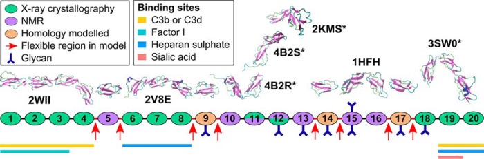
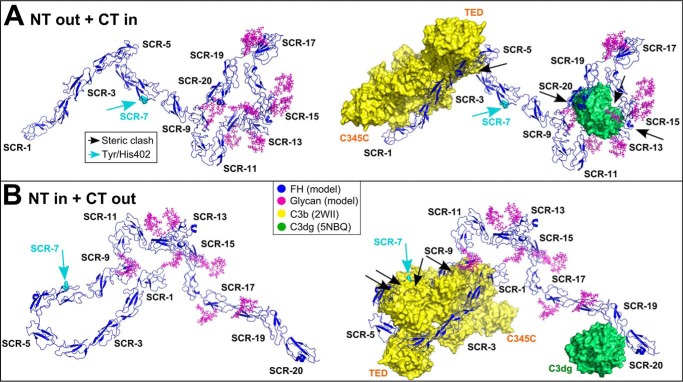
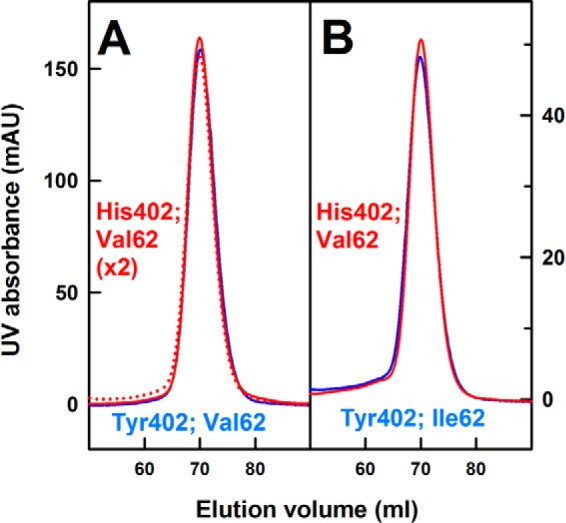
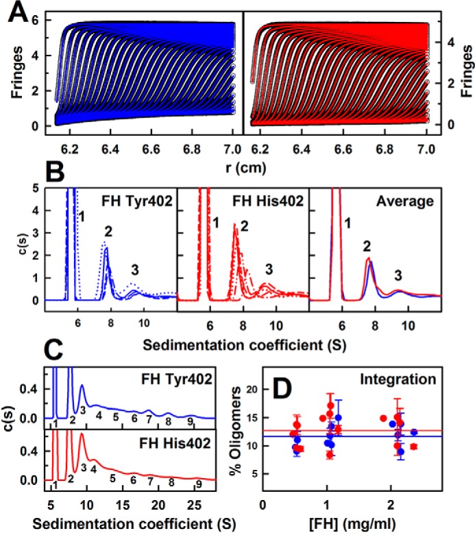
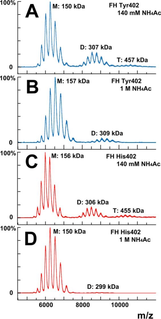
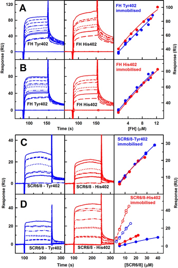
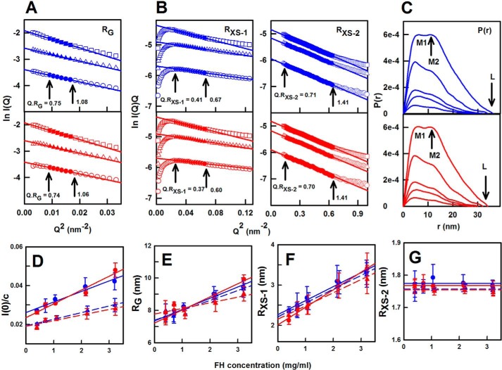
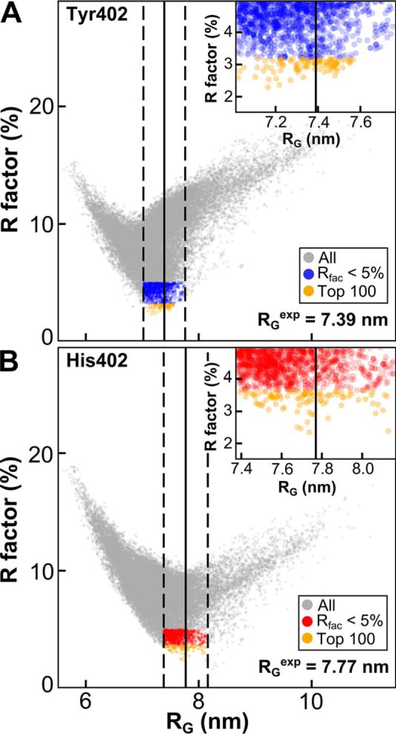
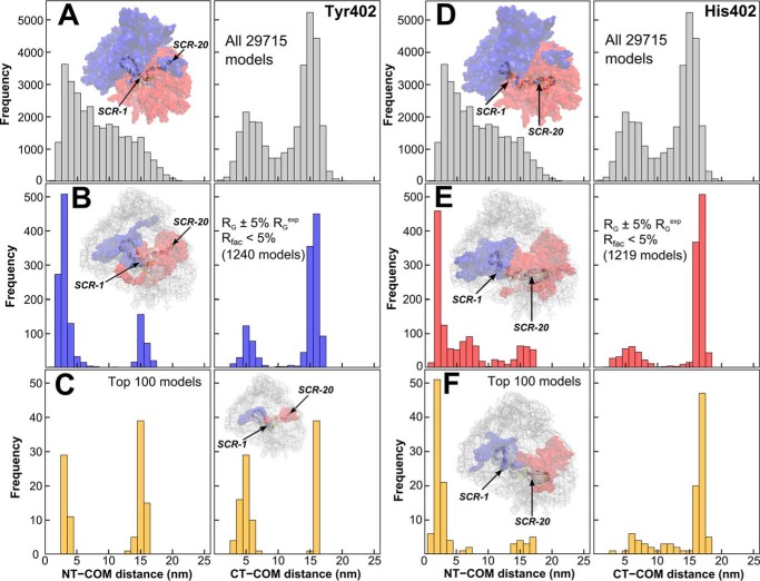
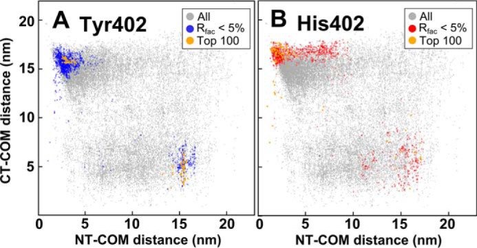
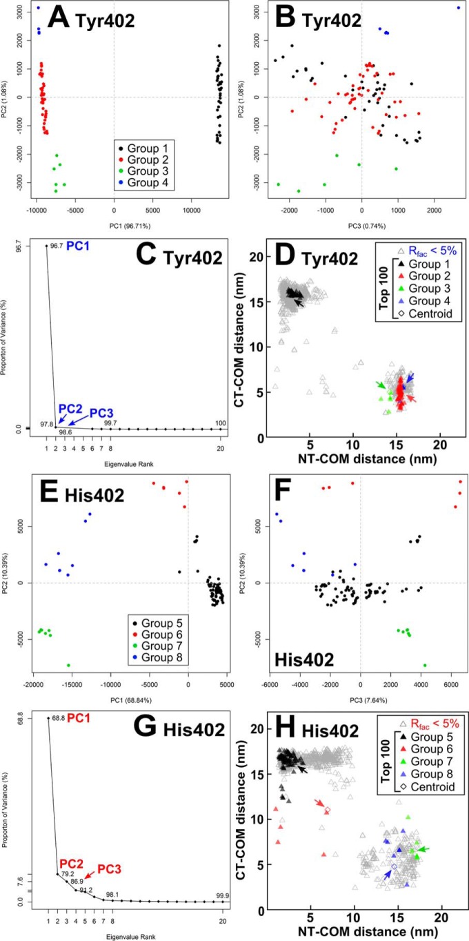
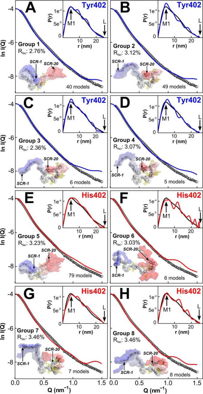
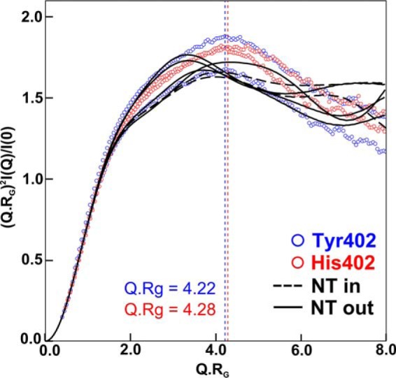
Factor H (FH) is the major regulator of C3b in the alternative pathway of the complement system in immunity. FH comprises 20 short complement regulator (SCR) domains, including eight glycans, and its Y402H polymorphism predisposes those who carry it to age-related macular degeneration. To better understand FH complement binding and self-association, we have studied the solution structures of both the His-402 and Tyr-402 FH allotypes. Analytical ultracentrifugation revealed that up to 12% of both FH allotypes self-associate, and this was confirmed by small-angle X-ray scattering (SAXS), MS, and surface plasmon resonance analyses. SAXS showed that monomeric FH has a radius of gyration ( ) of 7.2-7.8 nm and a length of 25 nm. Starting from known structures for the SCR domains and glycans, the SAXS data were fitted using Monte Carlo methods to determine atomistic structures of monomeric FH. The analysis of 29,715 physically realistic but randomized FH conformations resulted in 100 similar best-fit FH structures for each allotype. Two distinct molecular structures resulted that showed either an extended N-terminal domain arrangement with a folded-back C terminus or an extended C terminus and a folded-back N terminus. These two structures are the most accurate to date for glycosylated full-length FH. To clarify FH functional roles in host protection, crystal structures for the FH complexes with C3b and C3dg revealed that the extended N-terminal conformation accounted for C3b fluid-phase regulation, the extended C-terminal conformation accounted for C3d binding, and both conformations accounted for bivalent FH binding to glycosaminoglycans on the target cell surface.
因子 H(FH)是补体系统替代途径中 C3b 的主要调节剂。FH 由 20 个短补体调节(SCR)结构域组成,包括 8 个聚糖,其 Y402H 多态性使携带它的人易患年龄相关性黄斑变性。为了更好地理解 FH 补体结合和自组装,我们研究了 His-402 和 Tyr-402 FH 同种型的溶液结构。分析超速离心表明,多达 12%的 FH 同种型发生自组装,这通过小角度 X 射线散射(SAXS)、MS 和表面等离子体共振分析得到了证实。SAXS 表明,单体 FH 的回转半径( )为 7.2-7.8nm,长度为 25nm。从已知的 SCR 结构域和聚糖结构开始,使用蒙特卡罗方法对 SAXS 数据进行拟合,以确定单体 FH 的原子结构。对 29715 个物理上合理但随机的 FH 构象进行分析,得到了每个同种型的 100 个相似的最佳拟合 FH 结构。分析结果得到了两种截然不同的分子结构,一种是 N 端结构域伸展、C 端折叠的结构,另一种是 C 端伸展、N 端折叠的结构。这两种结构是迄今为止最准确的糖基化全长 FH 结构。为了阐明 FH 在宿主保护中的功能作用,FH 与 C3b 和 C3dg 的复合物晶体结构表明,伸展的 N 端构象解释了 C3b 液相调节,伸展的 C 端构象解释了 C3d 结合,两种构象都解释了二价 FH 与靶细胞表面糖胺聚糖的结合。