Dto. Química Física, Universidad Complutense de Madrid, Avenida Complutense s/n, 28040 Madrid, Spain; Instituto de Investigación Hospital Doce de Octubre (i+12), Avenida de Córdoba s/n, 28041 Madrid, Spain.
Dto. Química Física Biológica, Instituto de Química-Física "Rocasolano" (CSIC), Serrano 119, 28006 Madrid, Spain.
Biochim Biophys Acta Gen Subj. 2018 Dec;1862(12):2824-2834. doi: 10.1016/j.bbagen.2018.08.018. Epub 2018 Aug 30.
The fluorescent dye 10-N-nonyl acridine orange (NAO) is widely used as a mitochondrial marker. NAO was reported to have cytotoxic effects in cultured eukaryotic cells when incubated at high concentrations. Although the biochemical response of NAO-induced toxicity has been well identified, the underlying molecular mechanism has not yet been explored in detail.
We use optical techniques, including fluorescence confocal microscopy and lifetime imaging microscopy (FLIM) both in model membranes built up as giant unilamellar vesicles (GUVs) and cultured cells. These experiments are complemented with computational studies to unravel the molecular mechanism that makes NAO cytotoxic.
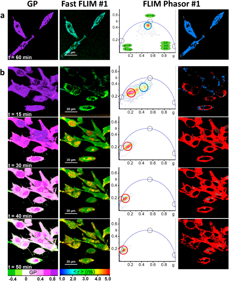
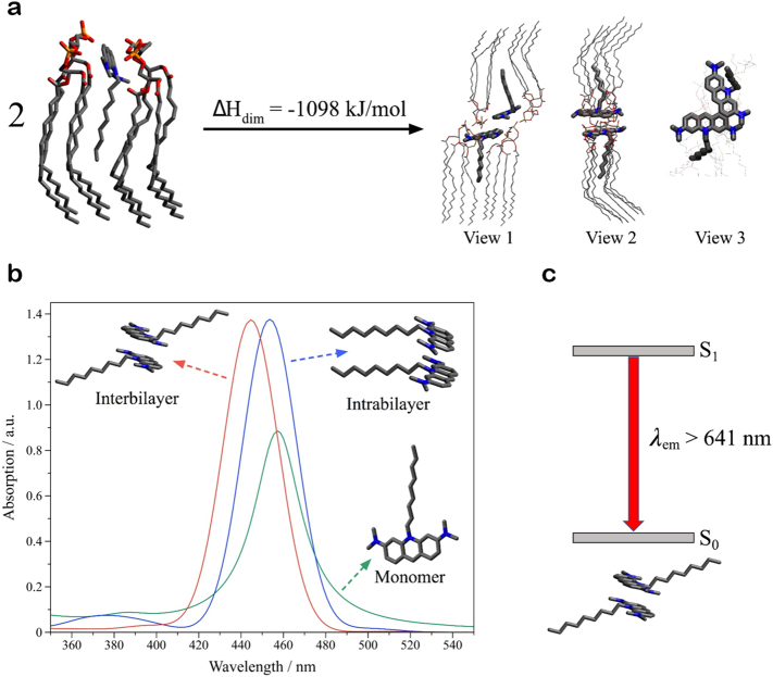
We have obtained direct evidence that NAO promotes strong membrane adhesion of negatively charged vesicles. The attractive forces are derived from van der Waals interactions between anti-parallel H-dimers of NAO molecules from opposing bilayers. Semi-empirical calculations have confirmed the supramolecular scenario by which anti-parallel NAO molecules form a zipper of bonds at the contact region. The membrane remodeling effect of NAO, as well as the formation of H-dimers, was also confirmed in cultured fibroblasts, as shown by the ultrastructure alteration of the mitochondrial cristae.
We conclude that membrane adhesion induced by NAO stacking accounts for the supramolecular basis of its cytotoxicity.
Mitochondria are a potential target for cancer and gene therapies. The alteration of the mitochondrial structure by membrane remodeling agents able to form supramolecular assemblies via adhesion properties could be envisaged as a new therapeutic strategy.
荧光染料 10-N-壬基吖啶橙(NAO)被广泛用作线粒体标记物。当在高浓度下孵育时,NAO 已被报道在培养的真核细胞中具有细胞毒性作用。尽管 NAO 诱导毒性的生化反应已经得到很好的鉴定,但尚未详细探索其潜在的分子机制。
我们使用光学技术,包括荧光共焦显微镜和寿命成像显微镜(FLIM),在模型膜(如巨单层囊泡(GUV)和培养细胞)中进行实验。这些实验辅以计算研究,以揭示使 NAO 具有细胞毒性的分子机制。
我们已经获得了直接证据,表明 NAO 促进带负电荷的囊泡的强烈膜粘附。吸引力来自于来自 opposing bilayers 的反平行 H-二聚体的范德华相互作用。半经验计算通过反平行 NAO 分子在接触区域形成键的拉链,证实了超分子方案。正如线粒体嵴的超微结构改变所表明的那样,NAO 对膜的重塑作用以及 H-二聚体的形成也在培养的成纤维细胞中得到了证实。
我们得出结论,由 NAO 堆积引起的膜粘附解释了其细胞毒性的超分子基础。
线粒体是癌症和基因治疗的潜在靶点。能够通过粘附特性形成超分子组装的膜重塑剂对线粒体结构的改变可以被设想为一种新的治疗策略。