Charité - Universitätsmedizin Berlin, corporate member of Freie Universität Berlin, Humboldt-Universität zu Berlin, and Berlin Institute of Health, Germany.
Center of Functionally Integrative Neuroscience, Clinical Institute, Aarhus University, Aarhus, Denmark.
PLoS One. 2018 Sep 26;13(9):e0202906. doi: 10.1371/journal.pone.0202906. eCollection 2018.
The purpose of this work is to investigate if the curve-fitting algorithm in Dynamic Contrast Enhanced (DCE) MRI experiments influences the diagnostic quality of calculated parameter maps.
We compared the Levenberg-Marquardt (LM) and a Bayesian method (BM) in DCE data of 42 glioma patients, using two compartmental models (extended Toft's and 2-compartment-exchange model). Logistic regression and an ordinal linear mixed model were used to investigate if the image quality differed between the curve-fitting algorithms and to quantify if image quality was affected for different parameters and algorithms. The diagnostic performance to discriminate between high-grade and low-grade gliomas was compared by applying a Wilcoxon signed-rank test (statistical significance p>0.05). Two neuroradiologists assessed different qualitative imaging features.
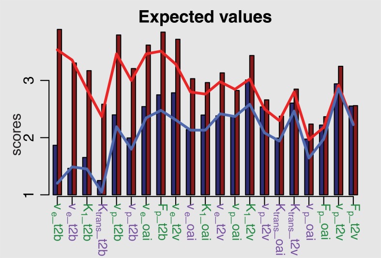
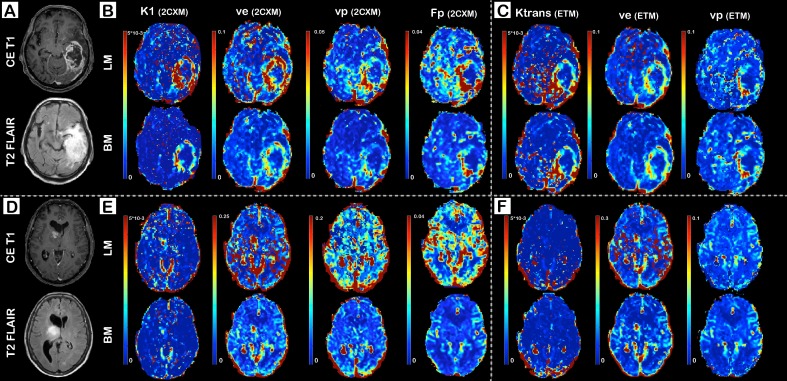
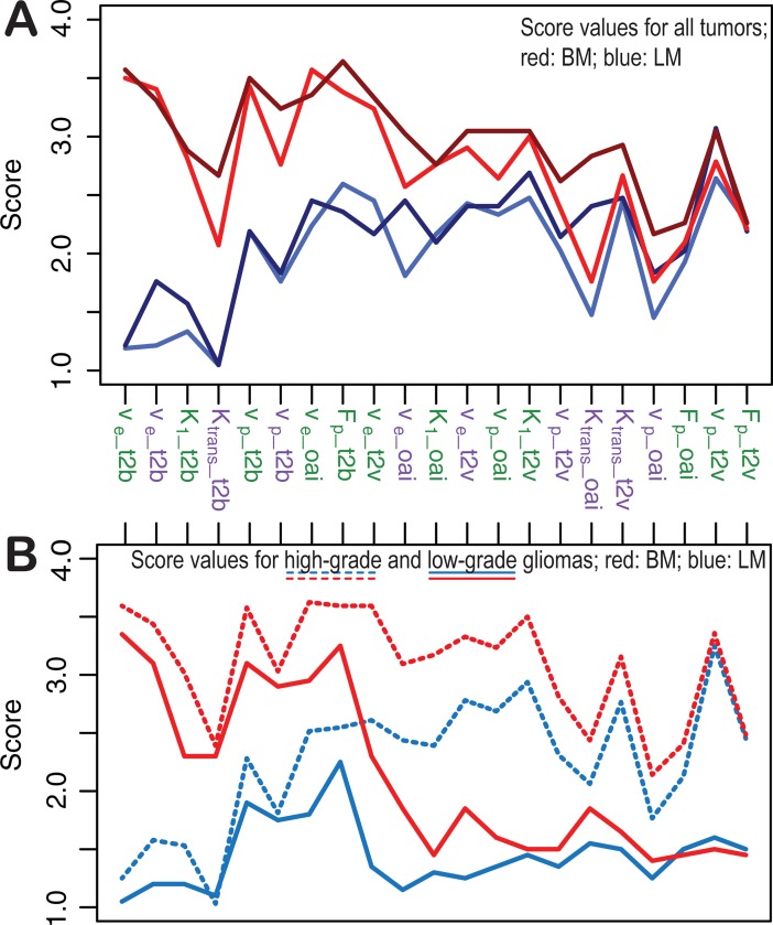
Parameter maps based on BM, particularly those describing the blood-brain barrier, were superior those based on LM. The image quality was found to be significantly improved (p<0.001) for BM when assessed through independent clinical scores. In addition, given a set of clinical scores, the generating algorithm could be predicted with high accuracy (area under the receiver operating characteristic curve between 0.91 and 1). Using linear mixed models, image quality was found to be improved when applying the 2-compartment-exchange model compared to the extended Toft's model, regardless of the underlying fitting algorithm. Tumor grades were only differentiated reliably on plasma volume maps when applying BM. The curve-fitting algorithm had, however, no influence on grading when using parameter maps describing the blood-brain barrier.
The Bayesian method has the potential to increase the diagnostic reliability of Dynamic Contrast Enhanced parameter maps in brain tumors. In our data, images based on the 2-compartment-exchange model were superior to those based on the extended Toft's model.
本研究旨在探讨动态对比增强磁共振成像(DCE MRI)实验中的曲线拟合算法是否会影响计算参数图的诊断质量。
我们比较了 Levenberg-Marquardt(LM)和贝叶斯(BM)两种方法在 42 例脑胶质瘤患者的 DCE 数据中的应用,使用了两种房室模型(扩展 Toft 模型和 2 房室交换模型)。采用逻辑回归和有序线性混合模型,研究曲线拟合算法之间的图像质量是否存在差异,并量化不同参数和算法对图像质量的影响。采用 Wilcoxon 符号秩检验比较两种算法对高级别和低级别胶质瘤的诊断效能(统计学意义 p>0.05)。两位神经放射科医生评估了不同的定性成像特征。
基于 BM 的参数图,尤其是描述血脑屏障的参数图,优于基于 LM 的参数图。独立的临床评分显示,基于 BM 的图像质量显著提高(p<0.001)。此外,在给定一组临床评分的情况下,生成算法可以被准确预测(受试者工作特征曲线下面积为 0.91 到 1 之间)。线性混合模型显示,与扩展 Toft 模型相比,使用 2 房室交换模型可以提高图像质量,与基础拟合算法无关。只有当使用 BM 时,才能可靠地根据血浆容积图区分肿瘤级别。然而,当使用描述血脑屏障的参数图时,曲线拟合算法对分级没有影响。
贝叶斯方法有可能提高脑肿瘤 DCE 参数图的诊断可靠性。在我们的数据中,基于 2 房室交换模型的图像优于基于扩展 Toft 模型的图像。