Antonacci Giuseppe, de Turris Valeria, Rosa Alessandro, Ruocco Giancarlo
Center for Life Nano Science@Sapienza, Istituto Italiano di Tecnologia, Rome, Italy.
Department of Biology and Biotechnology Charles Darwin, University of Rome"Sapienza", Rome, Italy.
Commun Biol. 2018 Sep 10;1:139. doi: 10.1038/s42003-018-0148-x. eCollection 2018.
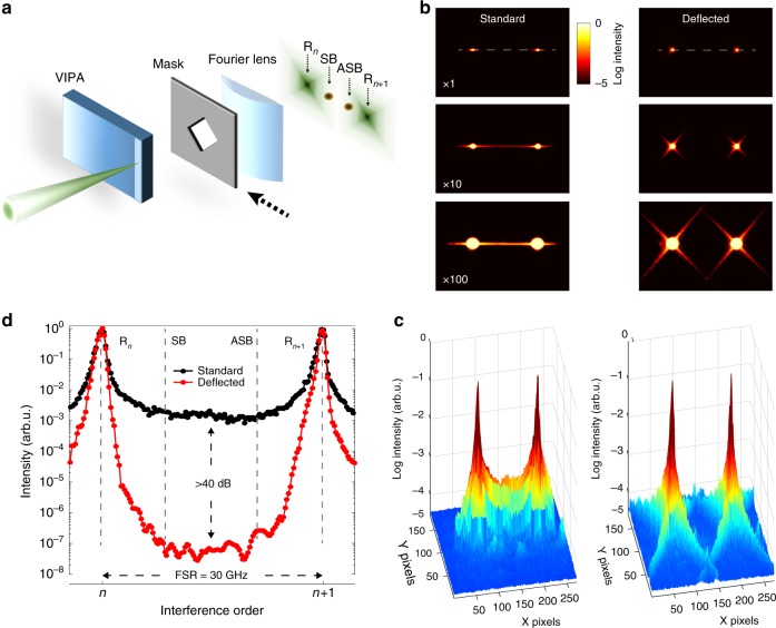
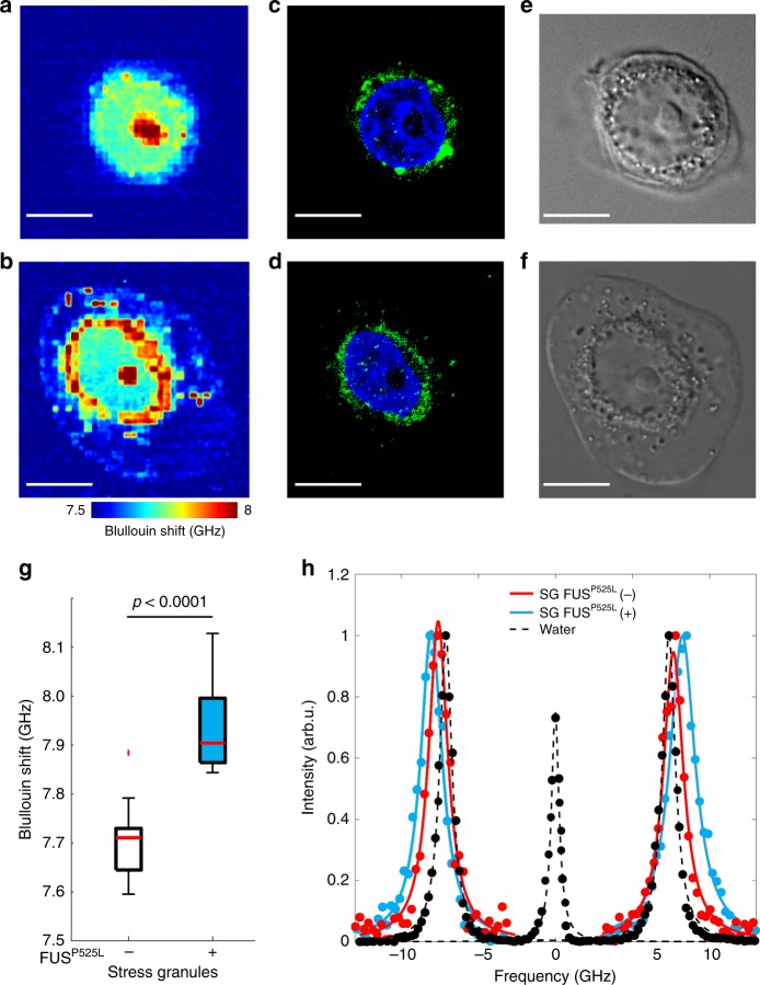
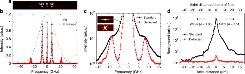
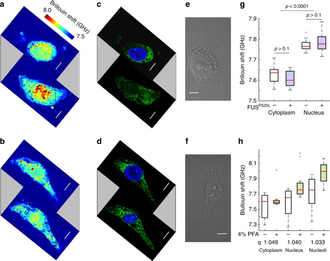
Altered cellular biomechanics have been implicated as key photogenic triggers in age-related diseases. An aberrant liquid-to-solid phase transition, observed in in vitro reconstituted droplets of FUS protein, has been recently proposed as a possible pathogenic mechanism for amyotrophic lateral sclerosis (ALS). Whether such transition occurs in cell environments is currently unknown as a consequence of the limited measuring capability of the existing techniques, which are invasive or lack of subcellular resolution. Here we developed a non-contact and label-free imaging method, named background-deflection Brillouin microscopy, to investigate the three-dimensional intracellular biomechanics at a sub-micron resolution. Our method exploits diffraction to achieve an unprecedented 10,000-fold enhancement in the spectral contrast of single-stage spectrometers, enabling, to the best of our knowledge, the first direct biomechanical analysis on intracellular stress granules containing ALS mutant FUS protein in fixed cells. Our findings provide fundamental insights on the critical aggregation step underlying the neurodegenerative ALS disease.
细胞生物力学改变被认为是与年龄相关疾病的关键发病诱因。最近,在体外重组的FUS蛋白液滴中观察到的异常液-固相变,被提出可能是肌萎缩侧索硬化症(ALS)的致病机制。由于现有技术的测量能力有限,这些技术要么具有侵入性,要么缺乏亚细胞分辨率,目前尚不清楚这种转变是否发生在细胞环境中。在这里,我们开发了一种非接触、无标记的成像方法,称为背景偏转布里渊显微镜,以亚微米分辨率研究三维细胞内生物力学。我们的方法利用衍射实现了单级光谱仪光谱对比度前所未有的10000倍增强,据我们所知,这使得首次对固定细胞中含有ALS突变FUS蛋白的细胞内应激颗粒进行直接生物力学分析成为可能。我们的研究结果为神经退行性ALS疾病背后的关键聚集步骤提供了基本见解。