Eunice Kennedy Shriver National Institute of Child Health and Human Development, National Institutes of Health, Bethesda, MD 20892.
Howard Hughes Medical Institute, Janelia Research Campus, Ashburn, VA 20147.
Proc Natl Acad Sci U S A. 2018 Oct 23;115(43):E10099-E10108. doi: 10.1073/pnas.1814552115. Epub 2018 Oct 4.
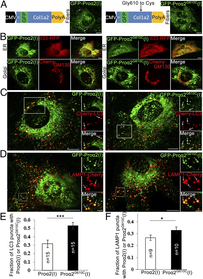
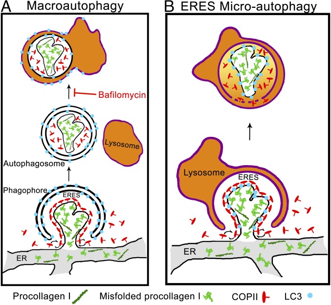
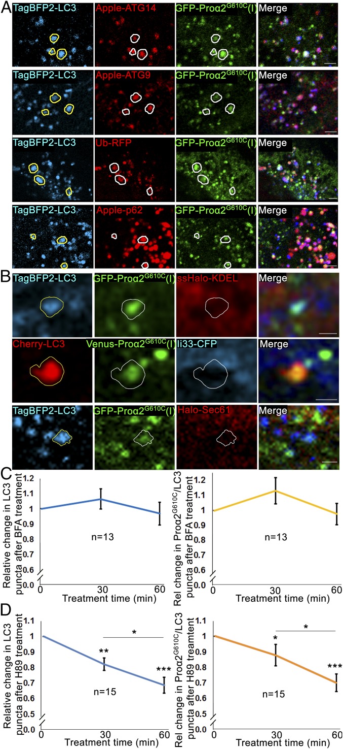
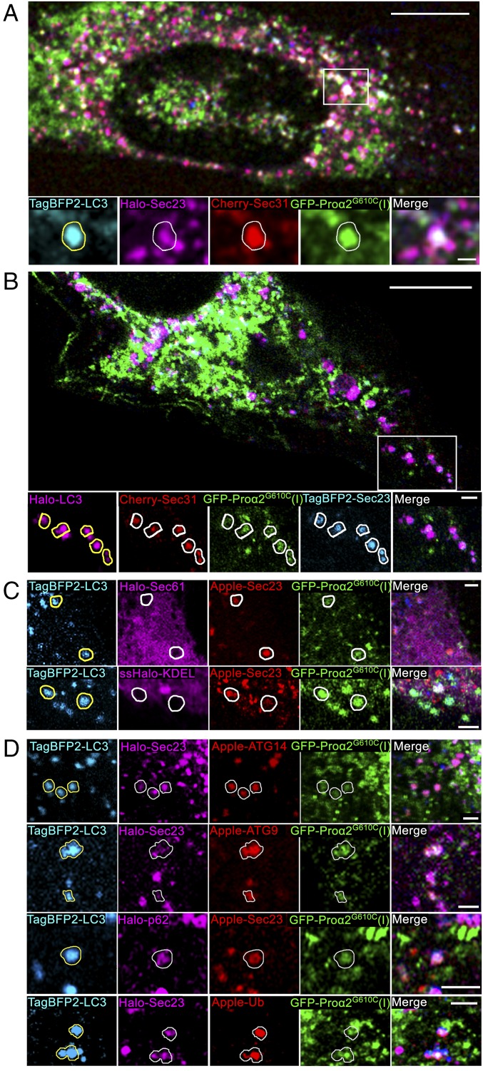
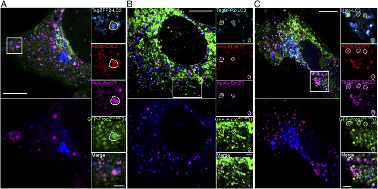
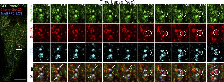
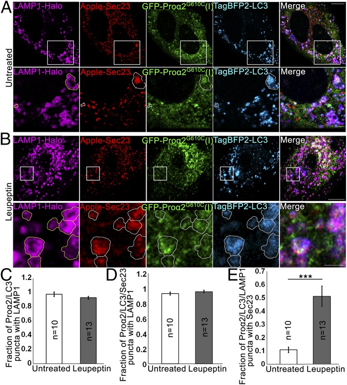
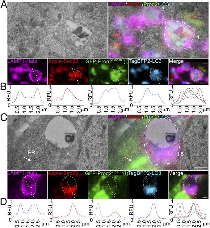
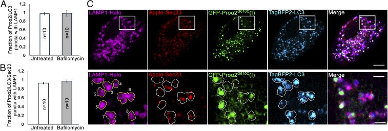
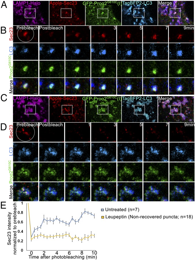
Type I collagen is the main component of bone matrix and other connective tissues. Rerouting of its procollagen precursor to a degradative pathway is crucial for osteoblast survival in pathologies involving excessive intracellular buildup of procollagen that is improperly folded and/or trafficked. What cellular mechanisms underlie this rerouting remains unclear. To study these mechanisms, we employed live-cell imaging and correlative light and electron microscopy (CLEM) to examine procollagen trafficking both in wild-type mouse osteoblasts and osteoblasts expressing a bone pathology-causing mutant procollagen. We found that although most procollagen molecules successfully trafficked through the secretory pathway in these cells, a subpopulation did not. The latter molecules appeared in numerous dispersed puncta colocalizing with COPII subunits, autophagy markers and ubiquitin machinery, with more puncta seen in mutant procollagen-expressing cells. Blocking endoplasmic reticulum exit site (ERES) formation suppressed the number of these puncta, suggesting they formed after procollagen entry into ERESs. The punctate structures containing procollagen, COPII, and autophagic markers did not move toward the Golgi but instead were relatively immobile. They appeared to be quickly engulfed by nearby lysosomes through a bafilomycin-insensitive pathway. CLEM and fluorescence recovery after photobleaching experiments suggested engulfment occurred through a noncanonical form of autophagy resembling microautophagy of ERESs. Overall, our findings reveal that a subset of procollagen molecules is directed toward lysosomal degradation through an autophagic pathway originating at ERESs, providing a mechanism to remove excess procollagen from cells.
I 型胶原蛋白是骨基质和其他结缔组织的主要成分。将其前胶原前体重新导向降解途径对于涉及细胞内前胶原过度积累的病理过程中破骨细胞的存活至关重要,这些前胶原折叠不正确和/或运输异常。这种重新定向的细胞机制尚不清楚。为了研究这些机制,我们采用活细胞成像和相关的光镜和电子显微镜(CLEM)来检查野生型小鼠成骨细胞和成骨细胞中前胶原的运输,这些成骨细胞表达一种导致骨病的突变前胶原。我们发现,尽管大多数前胶原分子在这些细胞中成功地通过分泌途径运输,但一部分没有。后者分子出现在许多分散的点状结构中,与 COPII 亚基、自噬标记物和泛素机器共定位,在突变型前胶原表达细胞中看到更多的点状结构。阻断内质网出口位点(ERES)的形成抑制了这些点状结构的数量,表明它们在前胶原进入 ERES 后形成。含有前胶原、COPII 和自噬标记物的点状结构不会向高尔基体移动,而是相对不动。它们似乎通过一种不依赖巴弗洛霉素的途径被附近的溶酶体迅速吞噬。CLEM 和光漂白后荧光恢复实验表明,吞噬作用是通过一种类似于 ERES 微自噬的非典型自噬形式发生的。总的来说,我们的研究结果表明,一部分前胶原分子通过起源于 ERES 的自噬途径被定向到溶酶体降解,为从细胞中去除多余的前胶原提供了一种机制。