Department of Anatomy & Embryology, Maastricht University, Maastricht, The Netherlands.
Department of Anatomy, Faculty of Science, Mahidol University, Bangkok, Thailand.
J Anat. 2018 Dec;233(6):724-739. doi: 10.1111/joa.12882. Epub 2018 Oct 7.
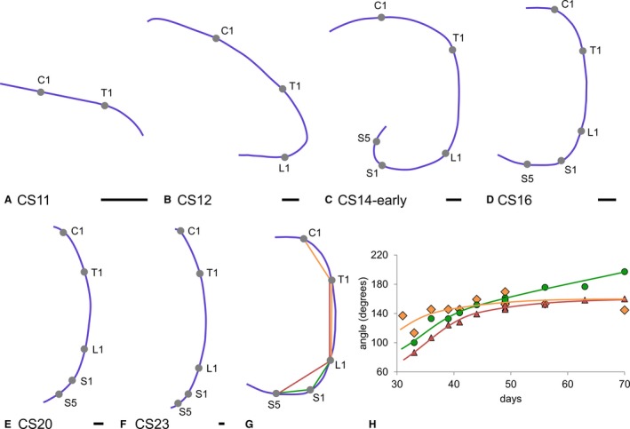
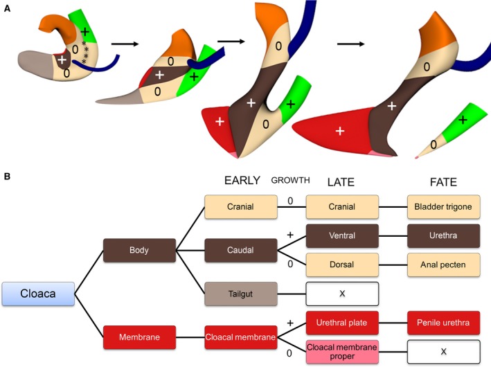
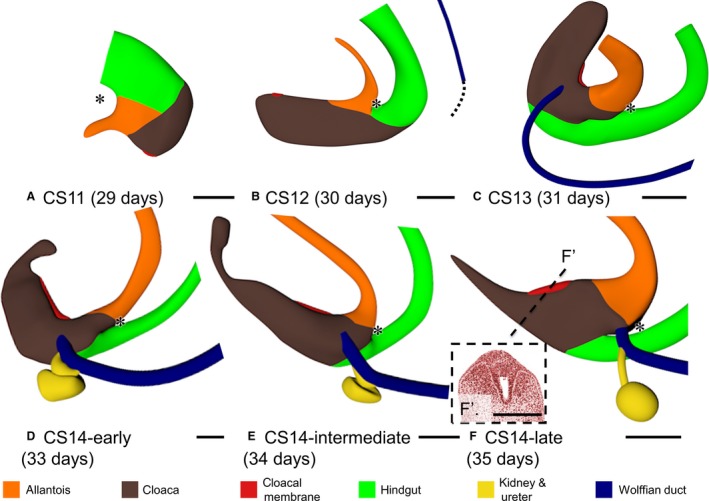
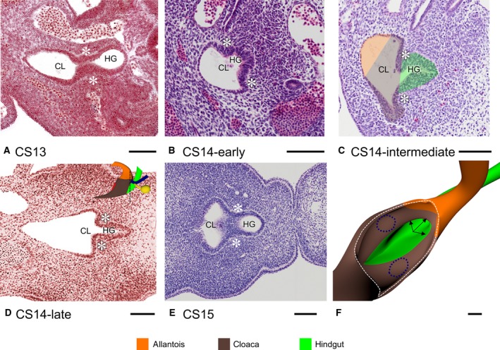
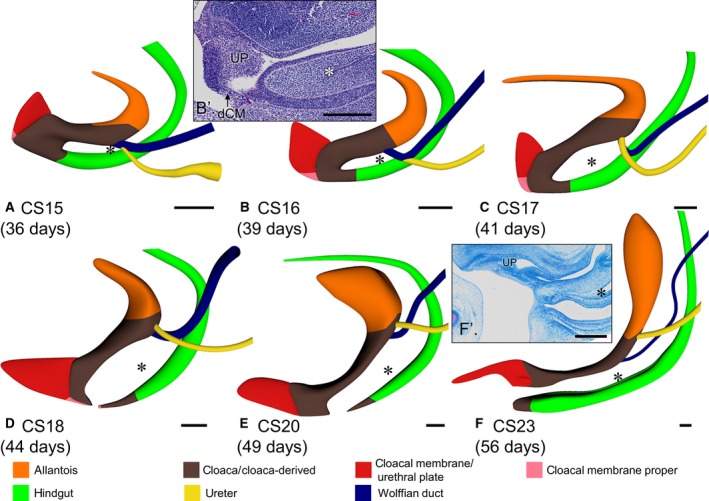
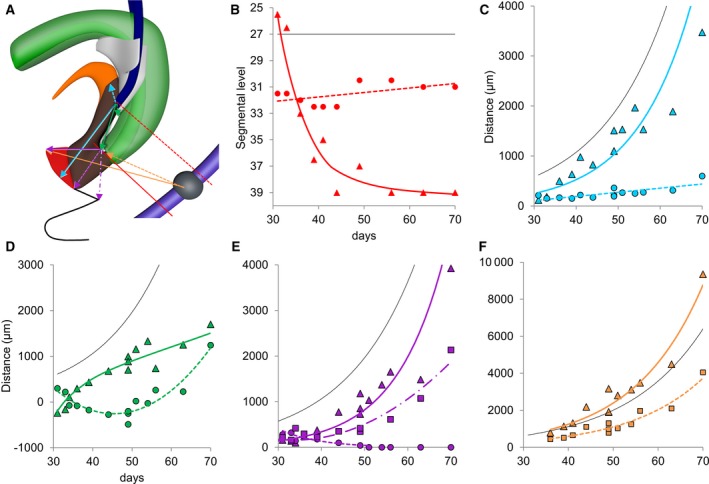
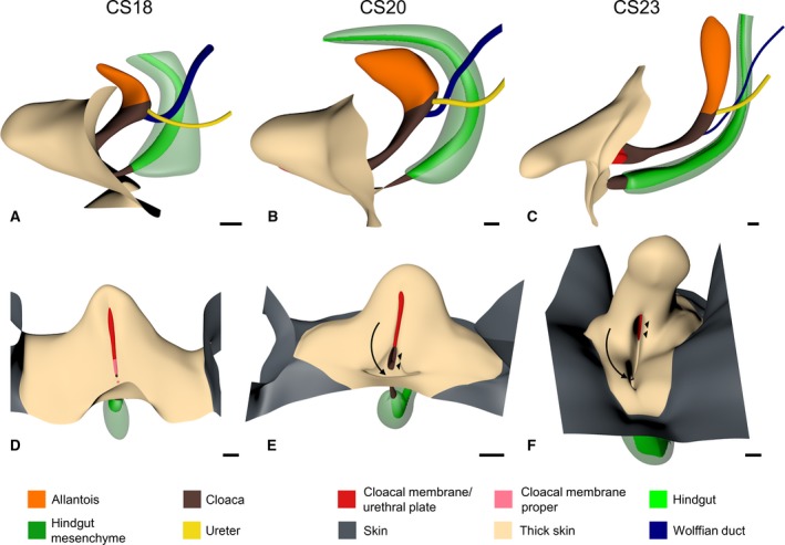
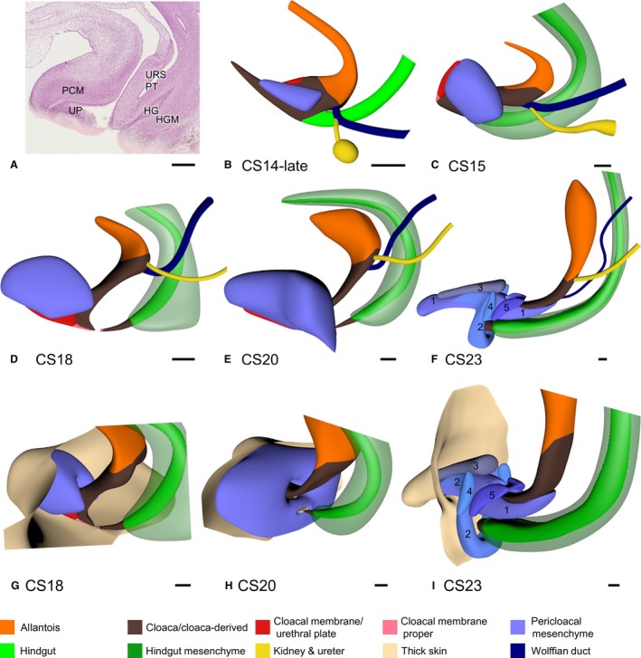
Subdivision of cloaca into urogenital and anorectal passages has remained controversial because of disagreements about the identity and role of the septum developing between both passages. This study aimed to clarify the development of the cloaca using a quantitative 3D morphological approach in human embryos of 4-10 post-fertilisation weeks. Embryos were visualised with Amira 3D-reconstruction and Cinema 4D-remodelling software. Distances between landmarks were computed with Amira3D software. Our main finding was a pronounced difference in growth between rapidly expanding central and ventral parts, and slowly or non-growing cranial and dorsal parts. The entrance of the Wolffian duct into the cloaca proved a stable landmark that remained linked to the position of vertebra S3. Suppressed growth in the cranial cloaca resulted in an apparent craniodorsal migration of the entrance of the Wolffian duct, while suppressed growth in the dorsal cloaca changed the entrance of the hindgut from cranial to dorsal on the cloaca. Transformation of this 'end-to-end' into an 'end-to-side' junction produced temporary 'lateral (Rathke's) folds'. The persistent difference in dorsoventral growth straightened the embryonic caudal body axis and concomitantly extended the frontally oriented 'urorectal (Tourneux's) septum' caudally between the ventral urogenital and dorsal anorectal parts of the cloaca. The dorsoventral growth difference also divided the cloacal membrane into a well-developed ventral urethral plate and a thin dorsal cloacal membrane proper, which ruptured at 6.5 weeks. The expansion of the pericloacal mesenchyme followed the dorsoventral growth difference and produced the genital tubercle. Dysregulation of dorsal cloacal development is probably an important cause of anorectal malformations: too little regressive development may result in anorectal agenesis, and too much regression in stenosis or atresia of the remaining part of the dorsal cloaca.
泄殖腔分为尿生殖和肛直肠两个通道,但由于人们对分隔这两个通道的中隔的性质和作用存在分歧,因此其划分一直存在争议。本研究旨在通过对受精后 4-10 周的人类胚胎进行定量的 3D 形态学研究,阐明泄殖腔的发育过程。使用 Amira 3D 重建和 Cinema 4D 重塑软件对胚胎进行可视化处理。使用 Amira3D 软件计算标志点之间的距离。我们的主要发现是,快速扩张的中央和腹侧部分与缓慢或不生长的颅侧和背侧部分之间的生长存在明显差异。沃尔夫管进入泄殖腔的入口被证明是一个稳定的标志,它与第 S3 节椎骨的位置相关联。颅侧泄殖腔生长受到抑制,导致沃尔夫管入口明显向颅侧迁移,而背侧泄殖腔生长受到抑制,使后肠入口从颅侧变为背侧。这种“端到端”的转变为“端到侧”的连接产生了临时的“侧(Rathke's)褶”。背腹向生长的持续差异使胚胎尾部的身体轴线变直,并使纵向的“尿生殖(Tourneux's)中隔”向后延伸,位于泄殖腔腹侧的泌尿生殖部和背侧的肛直肠部之间。背腹向生长差异还将泄殖腔膜分为发育良好的腹侧尿道板和薄的背侧泄殖腔膜,该膜在 6.5 周时破裂。泄殖腔周围中胚层的扩张遵循背腹向生长差异,并产生生殖结节。背侧泄殖腔发育的失调可能是肛直肠畸形的重要原因:发育不足可能导致肛直肠缺失,而发育过度则可能导致背侧泄殖腔剩余部分狭窄或闭锁。