Lomelino Carrie L, Andring Jacob T, McKenna Robert
University of Florida College of Medicine, Department of Biochemistry and Molecular Biology, Gainesville, FL 32610, USA.
Int J Med Chem. 2018 Sep 13;2018:9419521. doi: 10.1155/2018/9419521. eCollection 2018.
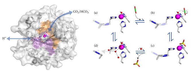
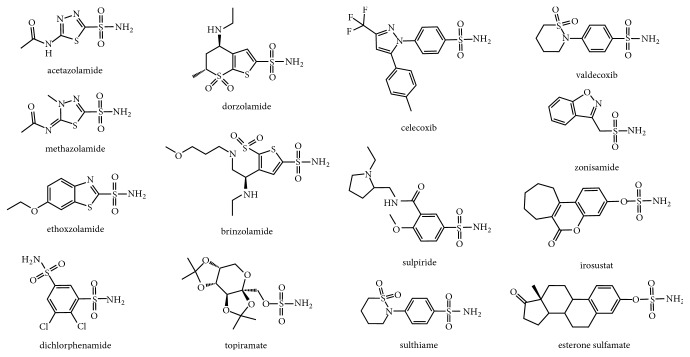
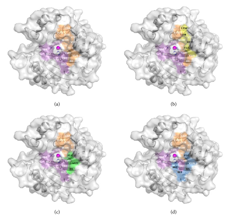
X-ray and neutron crystallography are powerful techniques utilized to study the structures of biomolecules. Visualization of enzymes in complex with substrate/product and the capture of intermediate states can be related to activity to facilitate understanding of the catalytic mechanism. Subsequent analysis of small molecule binding within the enzyme active site provides insight into mechanisms of inhibition, supporting the design of novel inhibitors using a structure-guided approach. The first X-ray crystal structures were determined for small, ubiquitous enzymes such as carbonic anhydrase (CA). CAs are a family of zinc metalloenzymes that catalyze the hydration of CO, producing HCO and a proton. The CA structure and ping-pong mechanism have been extensively studied and are well understood. Though the function of CA plays an important role in a variety of physiological functions, CA has also been associated with diseases such as glaucoma, edema, epilepsy, obesity, and cancer and is therefore recognized as a drug target. In this review, a brief history of crystallography and its impact on CA research is discussed.
X射线晶体学和中子晶体学是用于研究生物分子结构的强大技术。对与底物/产物结合的酶进行可视化以及捕捉中间状态,这与活性相关,有助于理解催化机制。随后对酶活性位点内小分子结合的分析,能深入了解抑制机制,支持采用结构导向方法设计新型抑制剂。最早测定的X射线晶体结构是针对诸如碳酸酐酶(CA)这类小型且普遍存在的酶。碳酸酐酶是一类锌金属酶,催化CO的水合作用,生成HCO和一个质子。碳酸酐酶的结构和乒乓机制已得到广泛研究且被充分理解。尽管碳酸酐酶的功能在多种生理功能中发挥重要作用,但它也与青光眼、水肿、癫痫、肥胖症和癌症等疾病有关,因此被视为一个药物靶点。在本综述中,将讨论晶体学的简要历史及其对碳酸酐酶研究的影响。