Universidade Federal do Rio de Janeiro, Centro de Ciências da Saúde, Instituto de Microbiologia Paulo de Góes, Departamento de Microbiologia Geral, Laboratório de Estudos Avançados de Microrganismos Emergentes e Resistentes, Rio de Janeiro, RJ, Brasil.
Universidade Federal do Rio de Janeiro, Centro de Ciências da Saúde, Instituto de Biofísica Carlos Chagas Filho, Laboratório de Biologia Celular de Fungos, Rio de Janeiro, RJ, Brasil.
Mem Inst Oswaldo Cruz. 2018 Oct 8;113(10):e180311. doi: 10.1590/0074-02760180311.
Scedosporium apiospermum is a ubiquitous, emerging and multidrug-resistant fungal pathogen with still rather unknown virulence mechanisms.
OBJECTIVES/METHODS: The cellular basis of the in vitro interaction between fungi and host cells/tissues is the determinant factor for the development of a successful in vivo infection. Herein, we evaluated the interaction of S. apiospermum conidia with lung epithelial (A549), lung fibroblast (MRC-5) and RAW 264.7 macrophages by light and scanning/transmission electron microscopy.
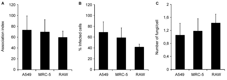
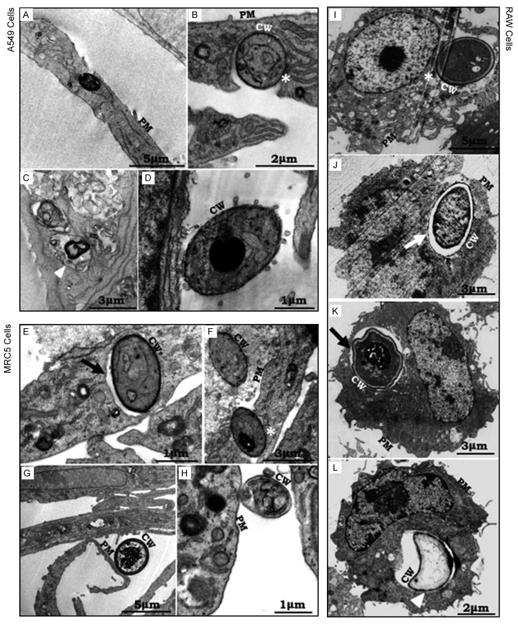
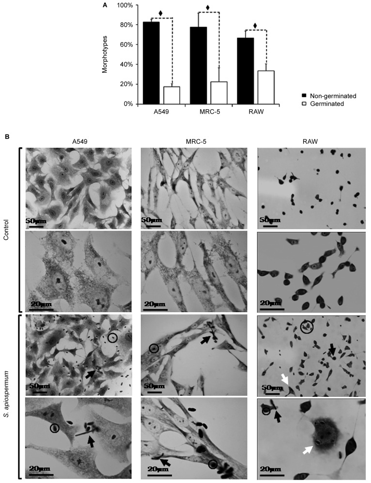
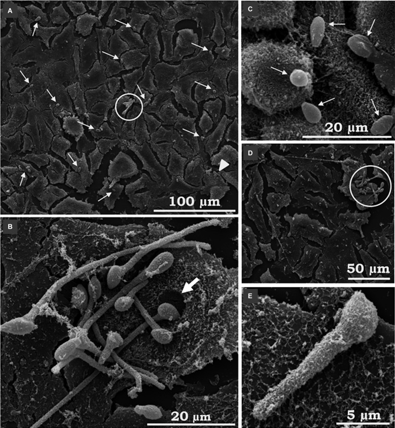
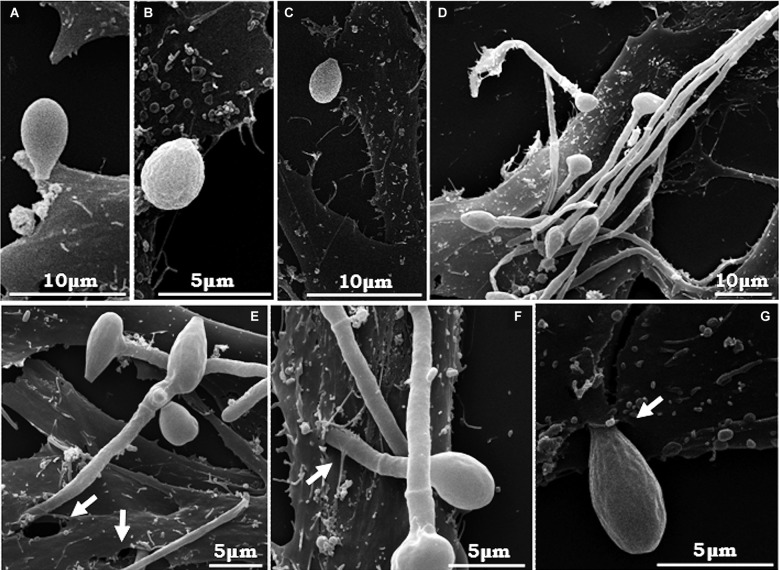
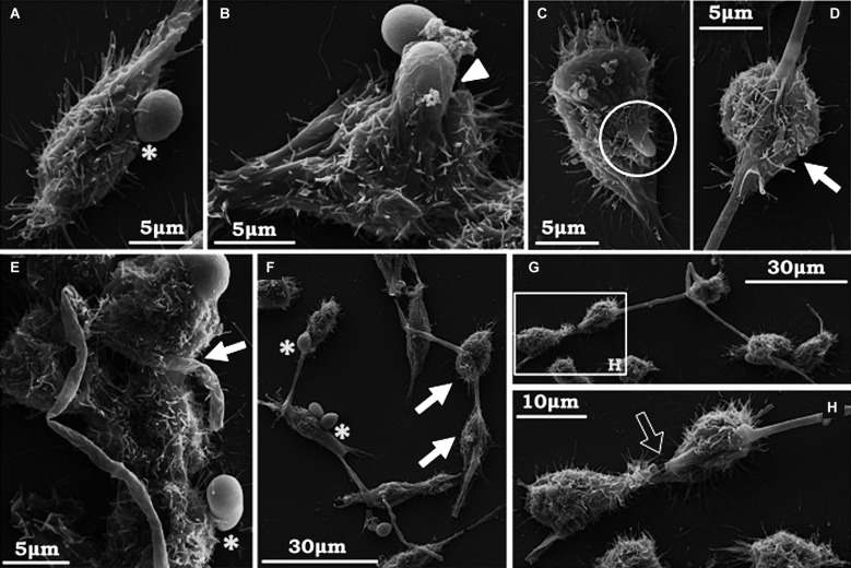
After 4 h of fungi-host cell contact, the percentage of infected mammalian cells and the number of fungi per infected cell was measured by light microscopy, and the following association indexes were calculated for A549, MRC-5 and macrophage cells: 73.2 ± 25.9, 69.7 ± 22.5 and 59.7 ± 11.1, respectively. Both conidia and germinated conidia were regularly observed interacting with the evaluated cells, with a higher prevalence of non-germinated conidia. Interestingly, nests of germinated conidia were evidenced at the surface of lung cells by scanning electron microscopy. Some germination projections and hyphae were seen penetrating/evading the mammalian cells. Furthermore, internalised conidia were seen within vacuoles as visualised by transmission electron microscopy.
The present study contributes to a better understanding of S. apiospermum pathogenesis by demonstrating the first steps of the infection process of this opportunistic fungus.
枝顶孢属(Scedosporium)是一种普遍存在、新兴的、多药耐药真菌病原体,其毒力机制仍知之甚少。
目的/方法:真菌与宿主细胞/组织之间体外相互作用的细胞基础是成功体内感染发展的决定因素。在此,我们通过光镜和扫描/透射电子显微镜评估了枝顶孢属分生孢子与肺上皮(A549)、肺成纤维细胞(MRC-5)和 RAW 264.7 巨噬细胞的相互作用。
在真菌与宿主细胞接触 4 小时后,通过光镜测量受感染哺乳动物细胞的百分比和每个受感染细胞中的真菌数量,并为 A549、MRC-5 和巨噬细胞计算以下关联指数:分别为 73.2±25.9、69.7±22.5 和 59.7±11.1。观察到规则的分生孢子和发芽的分生孢子与评估的细胞相互作用,未发芽的分生孢子更为常见。有趣的是,扫描电子显微镜显示在肺细胞表面有发芽的分生孢子巢。可以看到一些发芽的突起和菌丝穿透/逃避哺乳动物细胞。此外,透射电子显微镜显示内部化的分生孢子在空泡内。
本研究通过证明这种机会性真菌感染过程的最初步骤,有助于更好地了解枝顶孢属的发病机制。