Chondrou Vasiliki, Trochoutsou Katerina, Panayides Andreas, Efthimiou Maria, Stephanou Georgia, Demopoulos Nikos A
Division of Genetics, Cell and Developmental Biology, Department of Biology, University of Patras, 26 504, Patras, Greece.
J Biol Res (Thessalon). 2018 Oct 11;25:17. doi: 10.1186/s40709-018-0089-z. eCollection 2018 Dec.
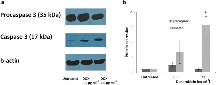
Doxorubicin is a widely used anticancer drug due to its broad spectrum of antitumor activity. Various mechanisms have been proposed for its cytostatic activity, including DNA intercalation, topoisomerase II inhibition, generation of free radicals and apoptosis. The present study aims to further clarify the cytostatic activity of doxorubicin by its specific effect on (a) DNA damage, (b) micronucleation and (c) apoptosis, using a combination of different methods and cell systems such as human lymphocytes and HL-60 human leukemic cells. DNA lesions were analyzed by the alkaline comet assay in combination with formamidopyrimidine (Fpg) and human 8-oxoguanine (hOGG1) repair enzymes. Micronucleation was investigated by the Cytokinesis-Block Micronucleus assay (CBMN) in combination with Fluorescence In Situ Hybridization analysis. Impairment on mitotic apparatus was investigated by double immunofluorescence of β- and γ-tubulin. Apoptotic cell frequency was determined by the CBMN cytome assay. Complementary to the above, caspase-3 level was investigated by Western blot.
It was found that doxorubicin generates DNA breakage induced by oxidative damage in DNA bases, which can be repaired by the Fpg and hOGG1 enzymes. Increased micronucleus frequency was identified mainly through chromosome breakage and, at a lesser extent, through chromosome delay. Analysis of mitotic spindle showed disturbance of chromosome orientation and centrosome duplication and/or separation, leading to aneuploidy. Enhanced frequency of apoptotic leukemic cells was also observed. Caspase-3 seems to be involved in the generation of apoptosis.
The aforementioned findings derived from different treatment schedules, doses and time of exposure on primary versus transformed cells extend our knowledge about doxorubicin genotoxicity and contribute to the better understanding of the mechanisms by which doxorubicin induces genotoxic effects on human cells.
由于阿霉素具有广泛的抗肿瘤活性,它是一种广泛使用的抗癌药物。关于其细胞生长抑制活性,已经提出了多种机制,包括DNA嵌入、拓扑异构酶II抑制、自由基生成和细胞凋亡。本研究旨在通过联合使用不同方法和细胞系统(如人淋巴细胞和HL-60人白血病细胞),研究阿霉素对(a)DNA损伤、(b)微核形成和(c)细胞凋亡的特定作用,进一步阐明其细胞生长抑制活性。通过碱性彗星试验结合甲酰胺嘧啶(Fpg)和人8-氧代鸟嘌呤(hOGG1)修复酶分析DNA损伤。通过胞质分裂阻滞微核试验(CBMN)结合荧光原位杂交分析研究微核形成。通过β-微管蛋白和γ-微管蛋白的双重免疫荧光研究对有丝分裂装置的损害。通过CBMN细胞分析法测定凋亡细胞频率。作为上述研究的补充,通过蛋白质免疫印迹法研究半胱天冬酶-3水平。
发现阿霉素会导致DNA碱基氧化损伤诱导的DNA断裂,这种损伤可由Fpg和hOGG1酶修复。微核频率增加主要是通过染色体断裂,在较小程度上是通过染色体延迟。有丝分裂纺锤体分析显示染色体定向以及中心体复制和/或分离受到干扰,导致非整倍体。还观察到白血病细胞凋亡频率增加。半胱天冬酶-3似乎参与了细胞凋亡的发生。
上述关于不同处理方案、剂量和暴露时间对原代细胞与转化细胞影响的研究结果,扩展了我们对阿霉素遗传毒性的认识,并有助于更好地理解阿霉素对人类细胞诱导遗传毒性作用的机制。