Yan Wei-Qiang, Xin Yong-Kang, Jing Yong, Li Gang-Feng, Wang Shu-Mei, Rong Wei-Cheng, Xiao Gang, Lei Xue-Bin, Li Bo, Hu Yu-Chuan, Cui Guang-Bin
From the Department of Radiology & Functional and Molecular Imaging Key Lab of Shaanxi Province, and.
Department of Pathology, Tangdu Hospital, the Military Medical University of PLA Airforce (Fourth Military Medical University), Shaanxi, PR China.
J Comput Assist Tomogr. 2018 Nov/Dec;42(6):873-880. doi: 10.1097/RCT.0000000000000800.
The aim of the study was to explore the efficacy of iodine quantification with dual-energy computed tomography (DECT) in differentiating thymoma, thymic carcinoma, and thymic lymphoma.
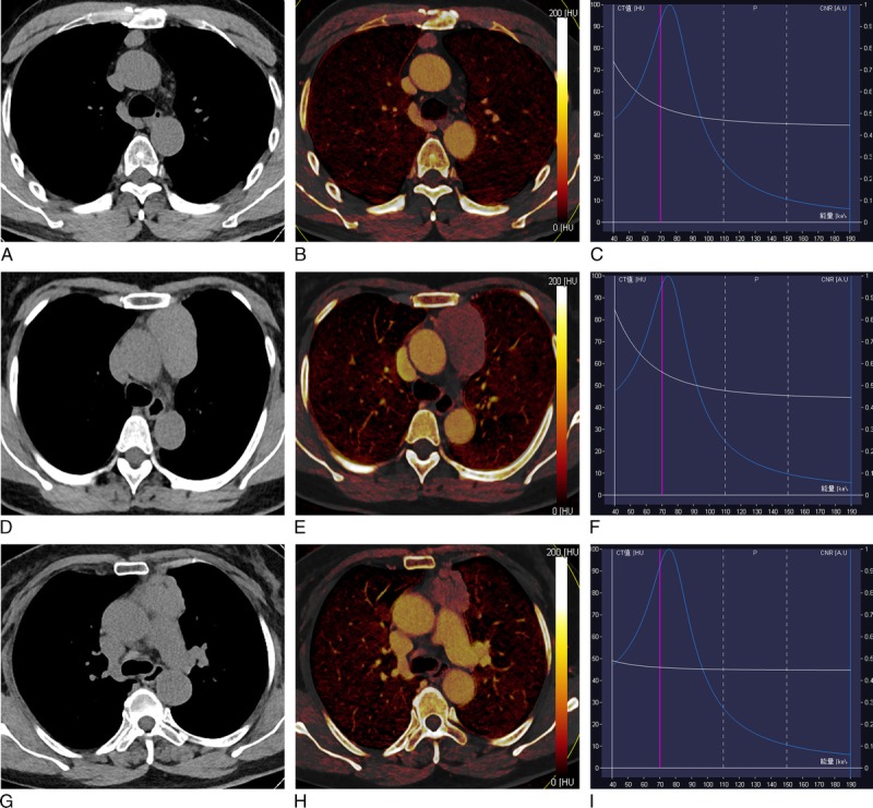
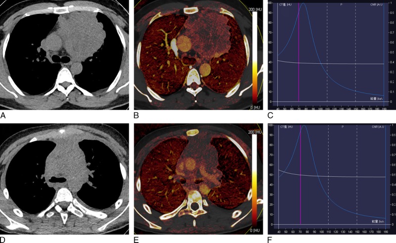
Fifty-seven patients with pathologically confirmed low-risk thymoma (n = 16), high-risk thymoma (n = 15), thymic carcinoma (n = 14), and thymic lymphoma (n = 12) underwent chest contrast-enhanced DECT scan were enrolled in this study. Tumor DECT parameters including iodine-related Hounsfield unit (IHU), iodine concentration (IC), mixed HU (MHU), and iodine ratio in dual phase, slope of energy spectral HU curve (λ), and virtual noncontrast (VNC) were compared for differences among 4 groups by one-way analysis of variance. Receiver operating characteristic curve was used to determine the efficacy for differentiating the low-risk thymoma from other thymic tumor by defined parameters.
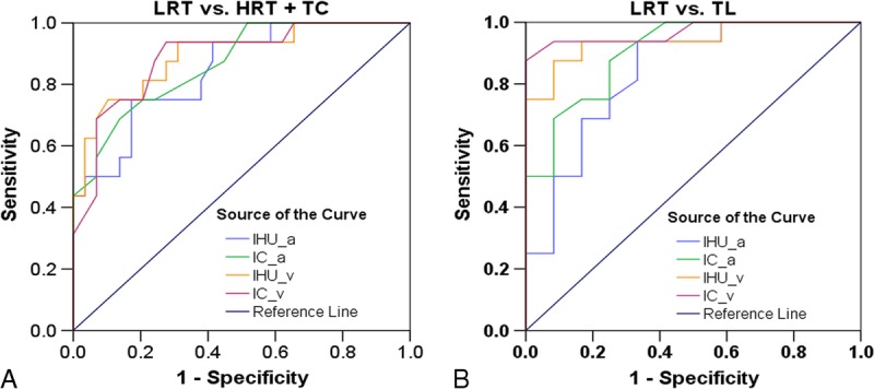
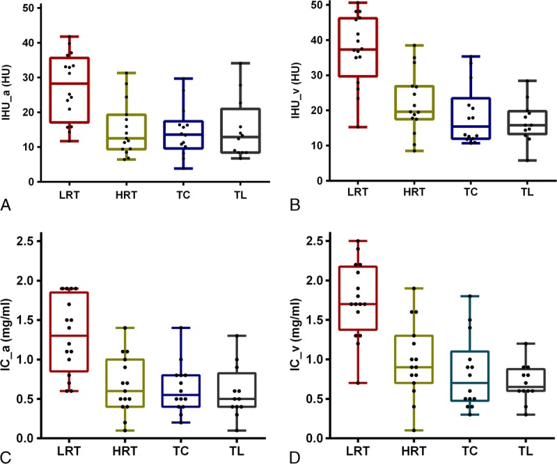
According to quantitative analysis, dual-phase IHU, IC, and MHU values in patients with low-risk thymoma were significantly increased compared with patients with high-risk thymoma, thymic carcinoma, and thymic lymphoma (P < 0.05/4).The venous phase IHU value yielded the highest performance with area under the curve of 0.893, 75.0% sensitivity, and 89.7% specificity for differentiating the low-risk thymomas from high-risk thymomas or thymic carcinoma at the cutoff value of 34.3 HU. When differentiating low-risk thymomas from thymic lymphoma, the venous phase IC value obtained the highest diagnostic efficacy with the area under the curve of 0.969, and sensitivity, specificity, and cutoff value were 87.5%, 100.0%, and 1.25 mg/mL, respectively.
Iodine quantification with DECT may be useful for differentiating the low-risk thymomas from other thymic tumors.
本研究旨在探讨双能计算机断层扫描(DECT)碘定量在鉴别胸腺瘤、胸腺癌和胸腺淋巴瘤中的疗效。
57例经病理证实的低危胸腺瘤(n = 16)、高危胸腺瘤(n = 15)、胸腺癌(n = 14)和胸腺淋巴瘤(n = 12)患者接受胸部对比增强DECT扫描并纳入本研究。通过单因素方差分析比较4组之间肿瘤DECT参数,包括碘相关亨氏单位(IHU)、碘浓度(IC)、混合HU(MHU)、双期碘比率、能谱HU曲线斜率(λ)和虚拟平扫(VNC)的差异。采用受试者操作特征曲线,通过定义的参数确定鉴别低危胸腺瘤与其他胸腺肿瘤的疗效。
根据定量分析,低危胸腺瘤患者的双期IHU、IC和MHU值与高危胸腺瘤、胸腺癌和胸腺淋巴瘤患者相比显著升高(P < 0.05/4)。静脉期IHU值表现最佳,曲线下面积为0.893,在截断值为34.3 HU时,鉴别低危胸腺瘤与高危胸腺瘤或胸腺癌的灵敏度为75.0%,特异度为89.7%。当鉴别低危胸腺瘤与胸腺淋巴瘤时,静脉期IC值诊断效能最高,曲线下面积为0.969,灵敏度、特异度和截断值分别为87.5%、100.0%和1.25 mg/mL。
DECT碘定量可能有助于鉴别低危胸腺瘤与其他胸腺肿瘤。