Department of Pathology, Amsterdam Neuroscience, Amsterdam UMC, VU University Medical Center, Amsterdam, The Netherlands.
Centre for Neuroscience and Trauma, Blizard Institute, Barts and the London School of Medicine and Dentistry, Queen Mary University of London, London, UK.
Neuropathol Appl Neurobiol. 2019 Aug;45(5):459-475. doi: 10.1111/nan.12525. Epub 2018 Nov 23.
Amyotrophic lateral sclerosis (ALS) is a chronic neurodegenerative disease characterized by progressive loss of motor neurons, muscle weakness, spasticity, paralysis and death usually within 2-5 years of onset. Neuroinflammation is a hallmark of ALS pathology characterized by activation of glial cells, which respond by upregulating small heat shock proteins (HSPBs), but the exact underlying pathological mechanisms are still largely unknown. Here, we investigated the association between ALS disease duration, lower motor neuron loss, TARDNA-binding protein 43 (TDP-43) pathology, neuroinflammation and HSPB expression.
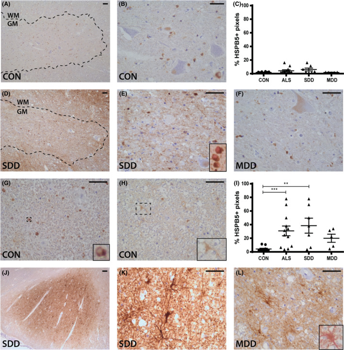
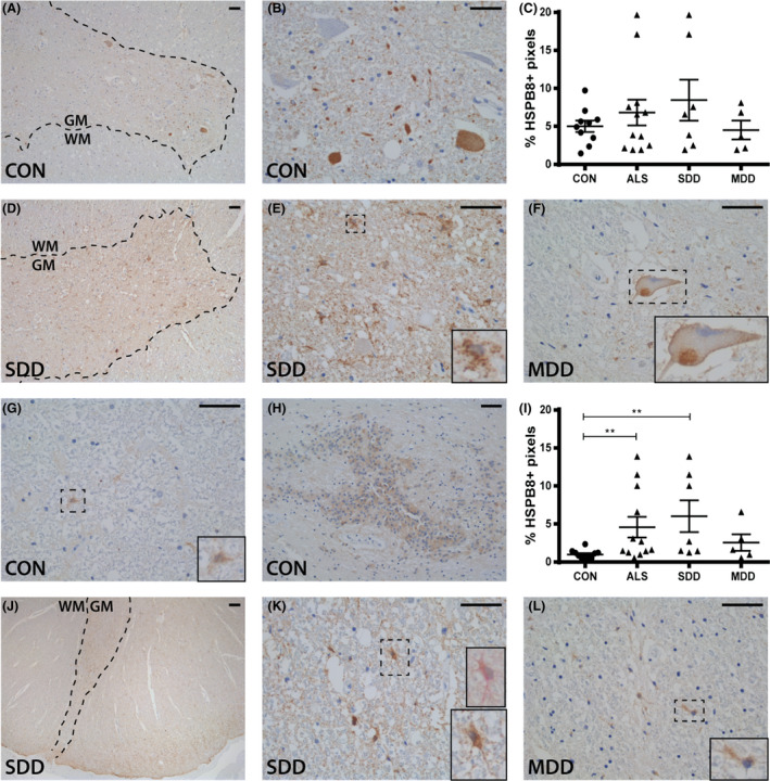
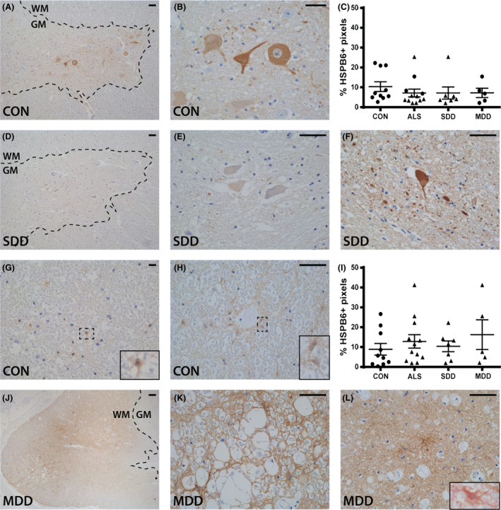
With immunohistochemistry, we examined HSPB1, HSPB5, HSPB6, HSPB8 and HSP16.2 expression in cervical, thoracic and sacral spinal cord regions in 12 ALS cases, seven with short disease duration (SDD), five with moderate disease duration (MDD), and ten age-matched controls. Expression was quantified using ImageJ to examine HSP expression, motor neuron numbers, microglial and astrocyte density and phosphorylated TDP-43 (pTDP-43+) inclusions.
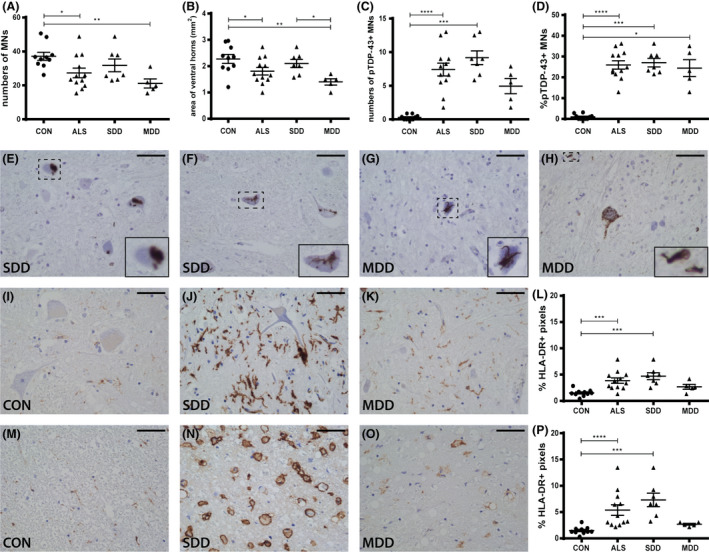
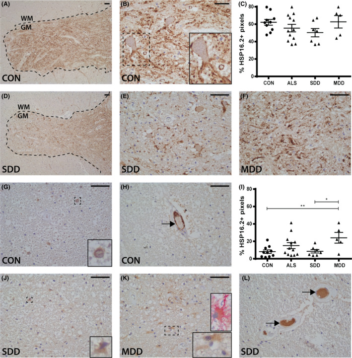
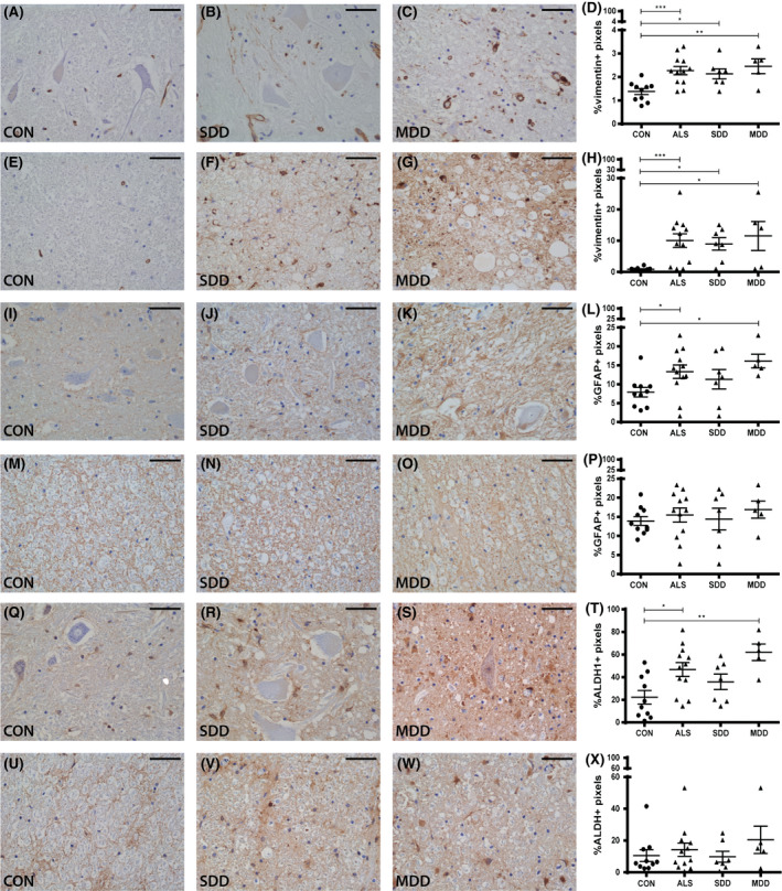
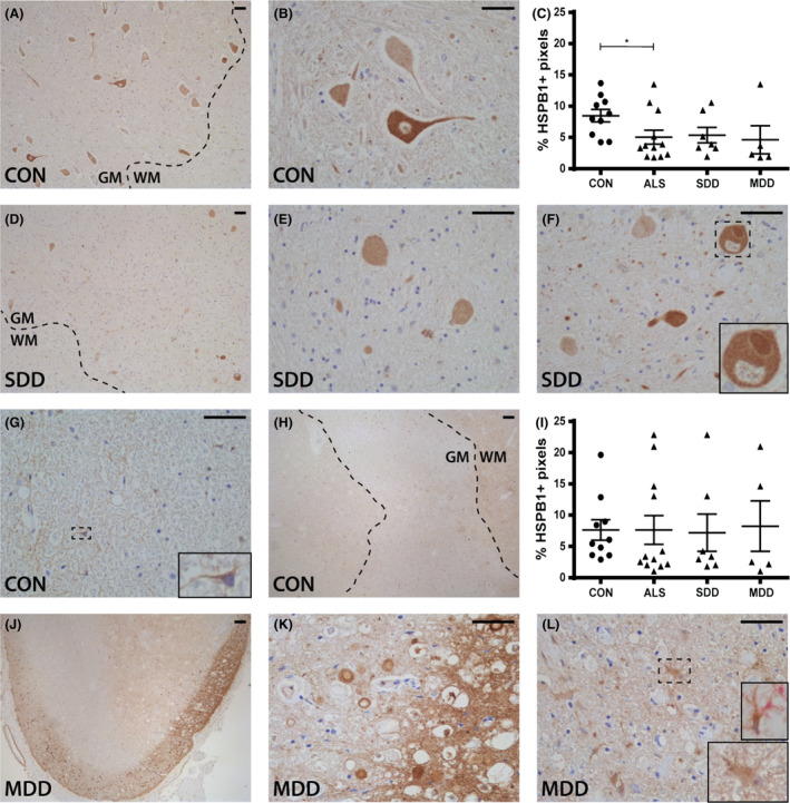
SDD was associated with elevated HSPB5 and 8 expression in lateral tract astrocytes, while HSP16.2 expression was increased in astrocytes in MDD cases. SDD cases had higher numbers of motor neurons and microglial activation than MDD cases, but similar levels of motor neurons with pTDP-43+ inclusions.
Increased expression of several HSPBs in lateral column astrocytes suggests that astrocytes play a role in the pathogenesis of ALS. SDD is associated with increased microgliosis, HSPB5 and 8 expression in astrocytes, and only minor changes in motor neuron loss. This suggests that the interaction between motor neurons, microglia and astrocytes determines neuronal fate and functional decline in ALS.
肌萎缩侧索硬化症(ALS)是一种慢性神经退行性疾病,其特征是运动神经元进行性丧失、肌肉无力、痉挛、瘫痪和死亡,通常在发病后 2-5 年内。神经炎症是 ALS 病理学的一个标志,其特征是神经胶质细胞的激活,这些细胞通过上调小热休克蛋白(HSPBs)来做出反应,但确切的潜在病理机制仍在很大程度上未知。在这里,我们研究了 ALS 疾病持续时间、下运动神经元丧失、TARDNA 结合蛋白 43(TDP-43)病理学、神经炎症和 HSPB 表达之间的关联。
我们使用免疫组织化学方法检查了 12 例 ALS 病例的颈、胸和骶骨脊髓区域中 HSPB1、HSPB5、HSPB6、HSPB8 和 HSP16.2 的表达,其中 7 例为疾病持续时间短(SDD),5 例为疾病持续时间中等(MDD),10 例为年龄匹配的对照组。使用 ImageJ 来检查 HSP 表达、运动神经元数量、小胶质细胞和星形胶质细胞密度以及磷酸化 TDP-43(pTDP-43+)包含物的表达来定量表达。
SDD 与侧束星形胶质细胞中 HSPB5 和 8 的表达升高有关,而 MDD 病例中 HSP16.2 在星形胶质细胞中的表达增加。SDD 病例的运动神经元数量和小胶质细胞激活高于 MDD 病例,但具有 pTDP-43+包含物的运动神经元数量相似。
几个 HSPB 在侧柱星形胶质细胞中的表达增加表明星形胶质细胞在 ALS 的发病机制中起作用。SDD 与小胶质细胞增多、星形胶质细胞中 HSPB5 和 8 的表达增加以及运动神经元丧失的微小变化有关。这表明运动神经元、小胶质细胞和星形胶质细胞之间的相互作用决定了 ALS 中神经元的命运和功能下降。