Department of Ophthalmology and Visual Sciences, School of Medicine, University of Alabama at Birmingham, Birmingham, Alabama, United States.
Invest Ophthalmol Vis Sci. 2018 Mar 20;59(4):AMD182-AMD194. doi: 10.1167/iovs.18-24883.
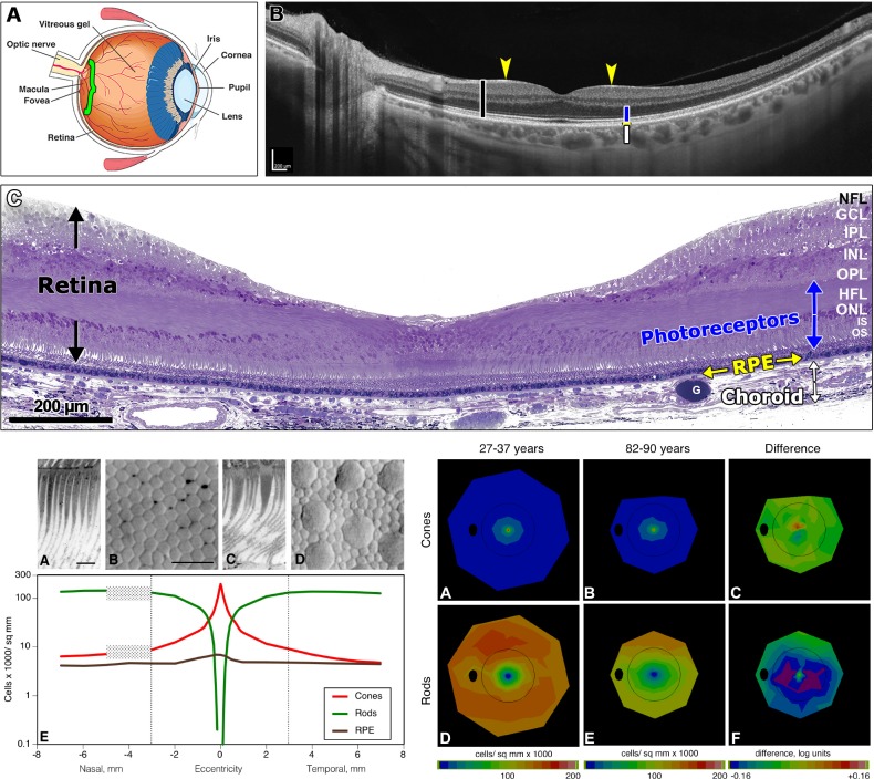
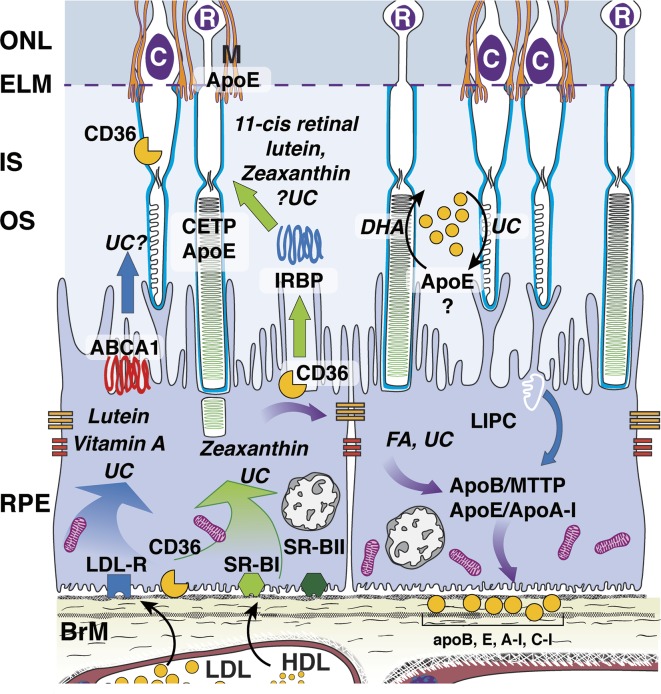
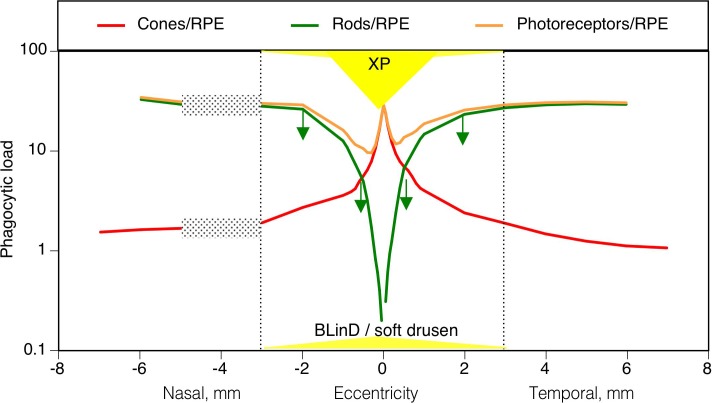
AMD pathobiology was irreversibly changed by the recent discovery of extracellular cholesterol-containing deposits in the subretinal space, between the photoreceptors and retinal pigment epithelium (RPE), called subretinal drusenoid deposits (SDDs). SDDs strikingly mirror the topography of rod photoreceptors in human macula, raising the question of whether an equivalent process results in a deposition related to foveal cones. Herein we propose that AMD's pathognomonic lesion-soft drusen and basal linear deposit (BLinD, same material, diffusely distributed)-is the leading candidate. Epidemiologic, clinical, and histologic data suggest that these deposits are most abundant in the central macula, under the fovea. Strong evidence presented in a companion article supports the idea that the dominant ultrastructural component is large apolipoprotein B,E-containing lipoproteins, constitutively secreted by RPE. Lipoprotein fatty acids are dominated by linoleate (implicating diet) rather than docosahexaenoate (implicating photoreceptors); we seek within the retina cellular relationships and dietary drivers to explain soft druse topography. The delivery of xanthophyll pigments to highly evolved and numerous Müller cells in the human fovea, through RPE, is one strong candidate, because Müller cells are the main reservoir of these pigments, which replenish from diet. We propose that the evolution of neuroglial relations and xanthophyll delivery that underlie exquisite human foveal vision came with a price, that is, soft drusen and sequela, long after our reproductive years.
AMD 的病理生物学发生了不可逆转的改变,最近在视网膜下空间(位于光感受器和视网膜色素上皮之间)发现了含有细胞外胆固醇的沉积物,称为视网膜下类 drusen 沉积物(SDD)。SDD 惊人地反映了人类黄斑中视杆光感受器的地形,这引发了一个问题,即是否存在同样的过程导致与中央凹锥细胞相关的沉积物。在此,我们提出 AMD 的特征性病变——软性玻璃膜疣和基底线性沉积物(BLinD,同一物质,弥漫分布)——是最主要的候选物。流行病学、临床和组织学数据表明,这些沉积物在黄斑中央最丰富,位于中央凹下。在一篇相关文章中提出的有力证据支持了这样一种观点,即主要的超微结构成分是由 RPE 持续分泌的大载脂蛋白 B、E 脂蛋白。脂蛋白中的脂肪酸以亚油酸(暗示饮食)为主,而不是二十二碳六烯酸(暗示光感受器);我们试图在视网膜中找到细胞关系和饮食驱动因素来解释软性玻璃膜疣的地形。叶黄素类色素通过 RPE 递送至人类黄斑中高度进化和众多的 Müller 细胞,这是一个强有力的候选物,因为 Müller 细胞是这些色素的主要储存库,它们可以从饮食中得到补充。我们提出,神经胶质关系和叶黄素类物质的传递是人类中央凹视力高度发达的基础,但也付出了代价,即软性玻璃膜疣和后遗症,在我们生殖期之后很久才出现。