Alhonen-Hongisto L, Leinonen P, Sinervirta R, Laine R, Winqvist R, Alitalo K, Jänne O A, Jänne J
Biochem J. 1987 Feb 15;242(1):205-10. doi: 10.1042/bj2420205.
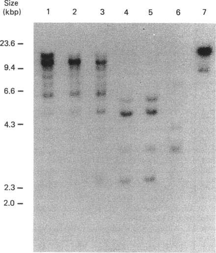
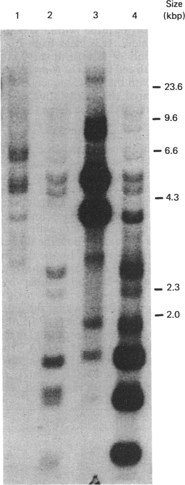
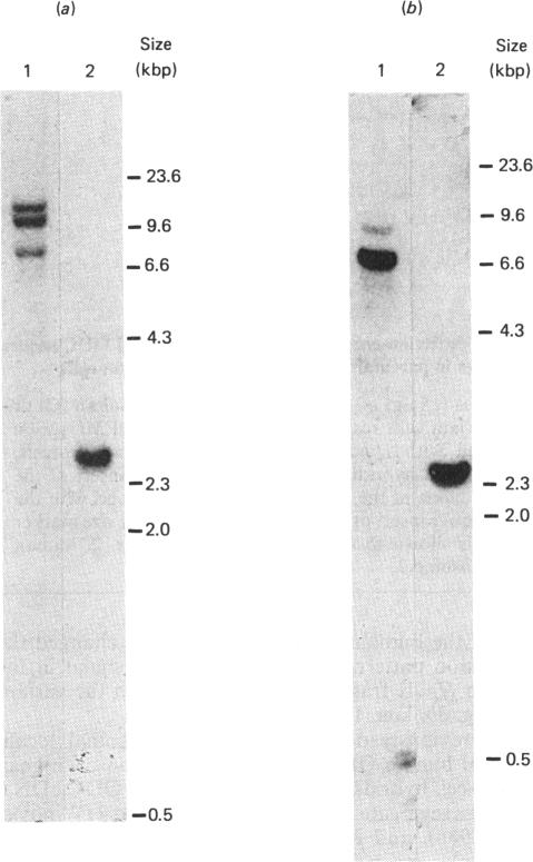
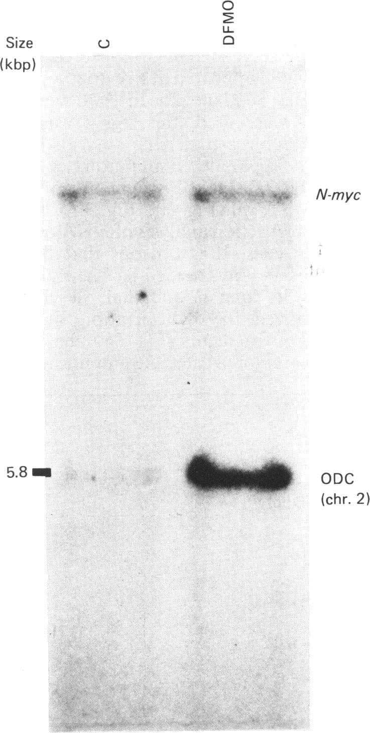
With the use of the isoschizomeric restriction endonucleases HpaII and MspI, we found that mouse tumour ornithine decarboxylase (ODC; EC 4.1.1.17) genes are extensively methylated. ODC genes in L1210 mouse leukaemia cells were apparently more methylated than in Ehrlich ascites carcinoma, as revealed by the use of HpaII endonuclease, yet the digestion of genomic DNA isolated from these two murine tumour cell lines with MspI, which cleaves at a CCGG sequence, also with internally methylated cytosine, resulted in an apparently identical restriction pattern. It is possible that the amplification of ODC genes in Ehrlich ascites-carcinoma cells in response to 2-difluoromethylornithine (DFMO) was associated with hypomethylation, or that less-methylated genes were amplified. A human myeloma (Sultan) cell line only revealed three separate hybridization signals when cleaved with HpaII. One of these signals was amplified under the pressure of DFMO. When cleaved with MspI, these three HpaII fragments disappeared and were replaced by a double signal of 2.3-2.4 kilobase-pairs (kbp) in size. The amplified ODC sequences in the Sultan myeloma cell line apparently originated from chromosome 2, as indicated by a unique hybridization signal in a 5.8 kbp HindIII fragment specific for the human ODC locus on chromosome 2. A comparison of different human cells, the Sultan myeloma, a lymphocytic B-cell leukaemia (Ball), normal mononuclear leucocytes and leucocytes obtained from leukaemia patients, revealed interesting differences in the methylation of ODC genes. The use of two restriction endonucleases (HpaII and CfoI), the cleavage site for both of which contains a CG sequence and which only cleave when cytosine is unmethylated, indicated that ODC genes in the lymphocytic leukaemia cells were much less methylated than those in the normal leucocytes or in the Sultan cells.
通过使用同裂酶限制性内切酶HpaII和MspI,我们发现小鼠肿瘤鸟氨酸脱羧酶(ODC;EC 4.1.1.17)基因被广泛甲基化。如使用HpaII内切酶所显示的,L1210小鼠白血病细胞中的ODC基因明显比艾氏腹水癌细胞中的甲基化程度更高,然而用MspI消化从这两种小鼠肿瘤细胞系中分离的基因组DNA,MspI在CCGG序列处切割,即使内部胞嘧啶甲基化时也能切割,结果产生了明显相同的限制性图谱。艾氏腹水癌细胞中ODC基因对2-二氟甲基鸟氨酸(DFMO)的反应性扩增可能与低甲基化有关,或者甲基化程度较低的基因被扩增了。一种人骨髓瘤(苏丹)细胞系在用HpaII切割时仅显示出三个单独的杂交信号。这些信号之一在DFMO的压力下被扩增。在用MspI切割时,这三个HpaII片段消失,取而代之的是一个大小为2.3 - 2.4千碱基对(kbp)的双重信号。苏丹骨髓瘤细胞系中扩增的ODC序列显然起源于2号染色体,这由2号染色体上人类ODC基因座特异性的5.8 kbp HindIII片段中的独特杂交信号所表明。对不同人类细胞的比较,即苏丹骨髓瘤、淋巴细胞B细胞白血病(鲍尔)、正常单核白细胞以及从白血病患者获得的白细胞,揭示了ODC基因甲基化方面有趣的差异。使用两种限制性内切酶(HpaII和CfoI),它们的切割位点都包含一个CG序列,并且只有在胞嘧啶未甲基化时才切割,这表明淋巴细胞白血病细胞中的ODC基因甲基化程度比正常白细胞或苏丹细胞中的低得多。