Xing Tiaosi, Hass Daniel T, Zhang Samuel S, Barnstable Colin J
Department of Anatomy and Cell Biology, East Carolina University, Greenville, NC, United States.
Department of Neural and Behavioral Sciences, Penn State College of Medicine, Hershey, PA, United States.
Front Cell Dev Biol. 2018 Oct 10;6:134. doi: 10.3389/fcell.2018.00134. eCollection 2018.
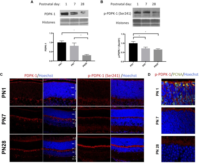
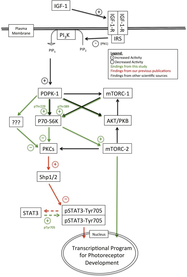
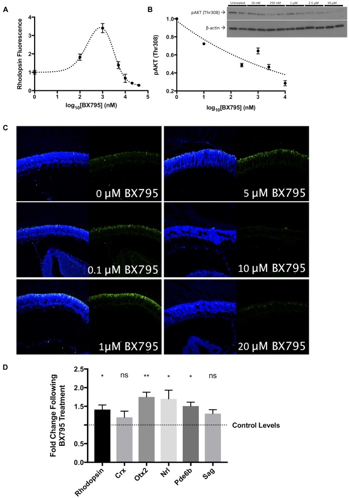
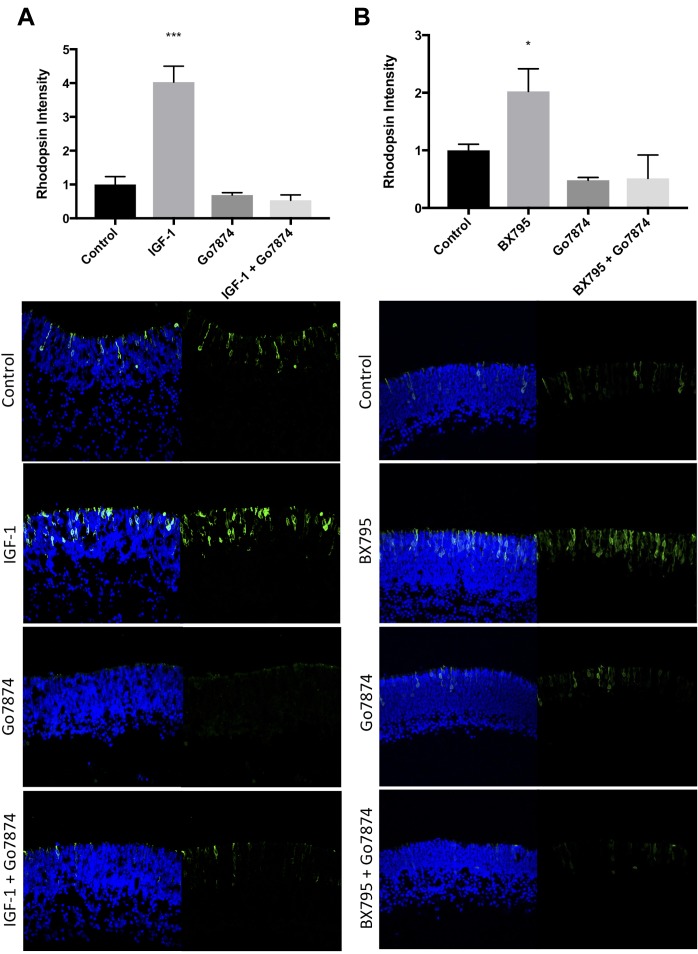
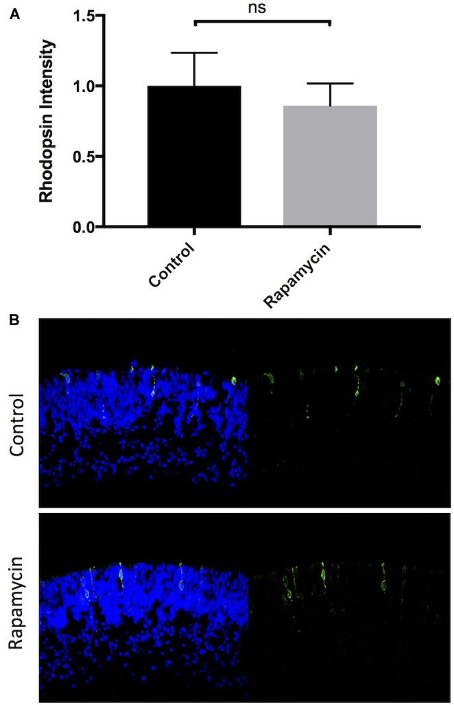
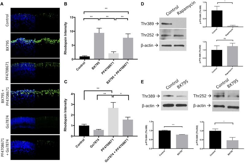
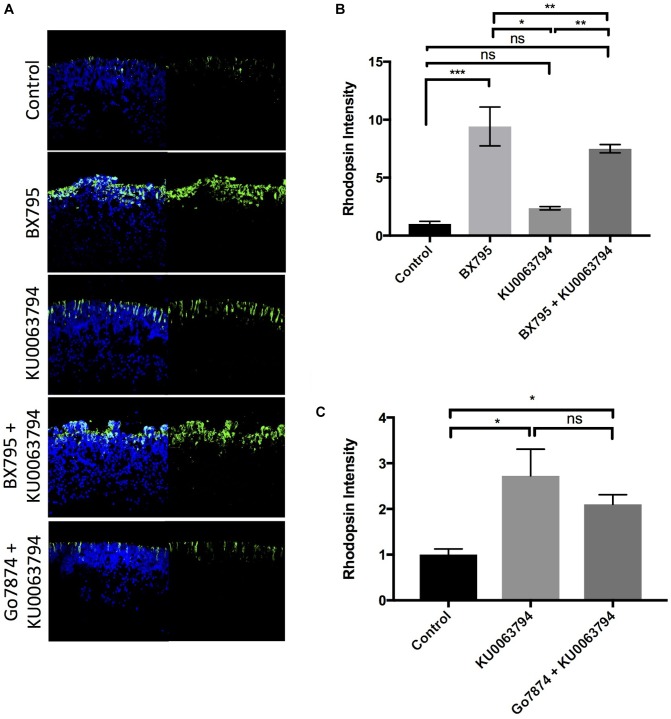
The transition of rod precursor cells to post-mitotic rod photoreceptors can be promoted by extrinsic factors such as insulin-like growth factor 1 (IGF-1), which regulates phosphatidylinositide concentration, and consequently the 3-phosphoinositide-dependent protein kinase-1 (PDPK-1). PDPK-1 is a 63 kDa cytoplasmic kinase that controls cell proliferation and differentiation. In the mouse retina, PDPK-1 and its phosphorylated derivative p-PDPK-1 (Ser241), showed peak expression during the first postnatal (PN) day with a substantial decline by PN7 and in the adult retina. Though initially widely distributed among cell types, PDPK-1 expression decreased first in the inner retina and later in the outer retina. When PDPK-1 is inhibited in neonatal retinal explants by BX795, there is a robust increase in rod photoreceptor numbers. The increase in rods depended on the activity of PKC, as BX795 had no effect when PKC is inhibited. Inhibition of PDPK-1-dependent kinases, such as P70-S6K, but not others, such as mTORC-1, stimulated rod development. The P70-S6K-dependent increase in rods appears to be correlated with phosphorylation of Thr252 and not at Thr389, a substrate of mTORC-1. This pathway is also inactive while PKC activity is inhibited. We also found that inhibition of the kinase mTORC-2, also stimulated by insulin activity, similarly increased rod formation, and this effect appears to be independent of PKC activity. This may represent a novel intracellular signaling pathway that also stimulates photoreceptor development. Consistent with previous studies, stimulation of STAT3 activity is sufficient to prevent any PDPK-1, P70-S6K, or mTORC2-dependent increase in rods. Together the data indicate that PDPK-1 and other intrinsic kinases downstream of IGF-1 are key regulators of rod photoreceptor formation.
杆状前体细胞向有丝分裂后杆状光感受器的转变可由诸如胰岛素样生长因子1(IGF-1)等外在因素促进,IGF-1调节磷脂酰肌醇浓度,进而调节3-磷酸肌醇依赖性蛋白激酶-1(PDPK-1)。PDPK-1是一种63 kDa的细胞质激酶,可控制细胞增殖和分化。在小鼠视网膜中,PDPK-1及其磷酸化衍生物p-PDPK-1(Ser241)在出生后第一天(PN)表达达到峰值,到PN7时大幅下降,在成年视网膜中表达量更低。尽管PDPK-1最初广泛分布于各种细胞类型中,但它在内视网膜中的表达首先下降,随后在外视网膜中下降。当用BX795抑制新生视网膜外植体中的PDPK-1时,杆状光感受器数量会显著增加。杆状光感受器数量的增加依赖于蛋白激酶C(PKC)的活性,因为当PKC被抑制时,BX795没有效果。抑制PDPK-1依赖性激酶,如P70-S6K,但不抑制其他激酶,如mTORC-1,可刺激杆状光感受器的发育。P70-S6K依赖性的杆状光感受器数量增加似乎与Thr252的磷酸化有关,而与mTORC-1的底物Thr389的磷酸化无关。当PKC活性被抑制时,该途径也无活性。我们还发现,抑制同样受胰岛素活性刺激的激酶mTORC-2,同样会增加杆状光感受器的形成,而且这种效应似乎与PKC活性无关。这可能代表了一种新的细胞内信号通路,也能刺激光感受器的发育。与之前的研究一致,刺激信号转导和转录激活因子3(STAT3)的活性足以阻止任何由PDPK-1、P70-S6K或mTORC2介导的杆状光感受器数量增加。这些数据共同表明,PDPK-1以及IGF-1下游的其他内在激酶是杆状光感受器形成的关键调节因子。