German Cancer Consortium (Deutsches Konsortium für Translationale Krebsforschung), Heidelberg, Germany.
Georg-Speyer-Haus, Institute for Tumor Biology and Experimental Therapy, Frankfurt am Main, Germany.
Cell Mol Gastroenterol Hepatol. 2018 Aug 14;6(4):477-493.e1. doi: 10.1016/j.jcmgh.2018.08.001. eCollection 2018.
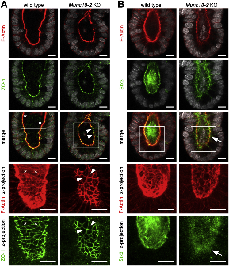
BACKGROUND & AIMS: Microvillus inclusion disease (MVID) is a congenital intestinal malabsorption disorder caused by defective apical vesicular transport. Existing cellular models do not fully recapitulate this heterogeneous pathology. The aim of this study was to characterize 3-dimensional intestinal organoids that continuously generate polarized absorptive cells as an accessible and relevant model to investigate MVID.
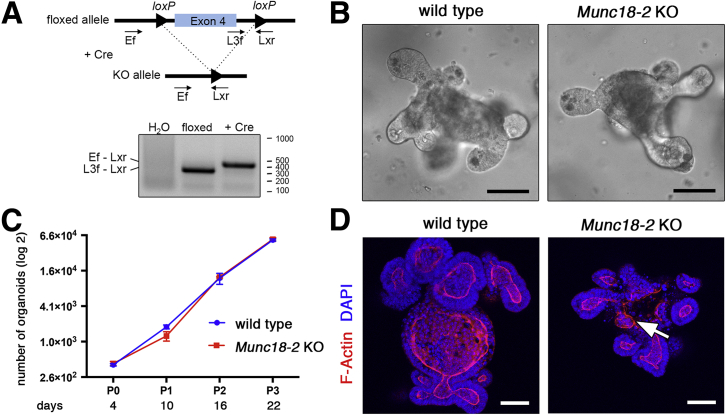
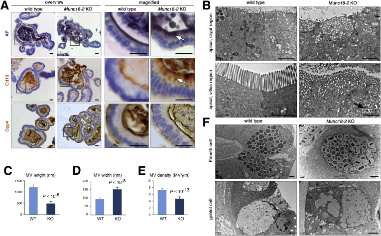
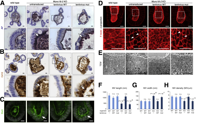
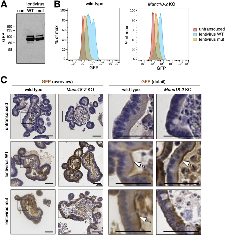
Intestinal organoids from /-null mice that are deficient for apical vesicular transport were subjected to enterocyte-specific differentiation protocols. Lentiviral rescue experiments were performed using human MUNC18-2 variants. Apical trafficking and microvillus formation were characterized by confocal and transmission electron microscopy. Spinning disc time-lapse microscopy was used to document the lifecycle of microvillus inclusions.
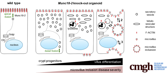
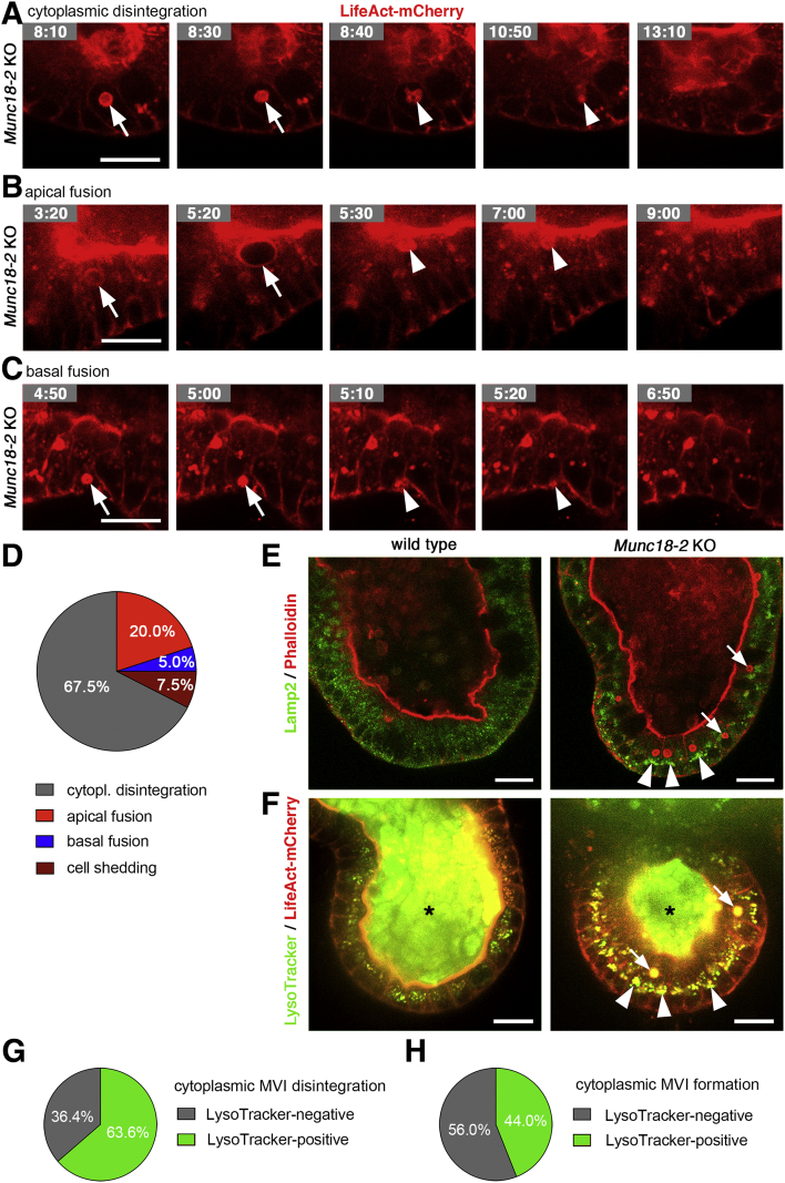
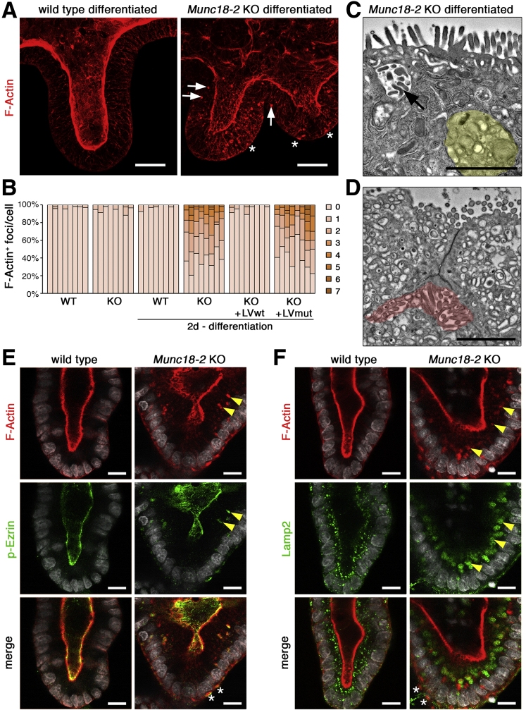
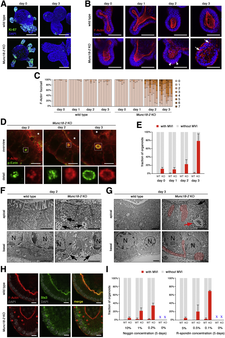
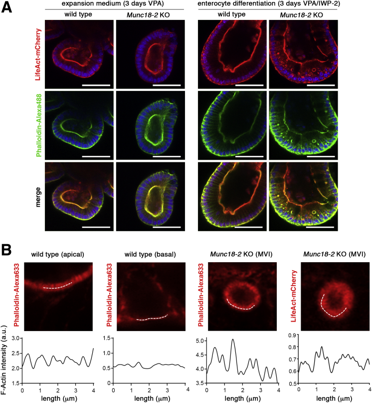
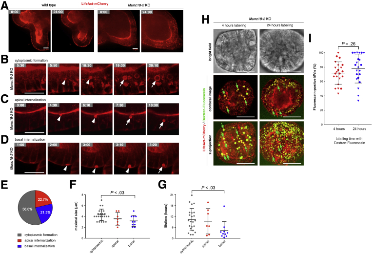
Loss of / recapitulated the pathologic features observed in patients with MUNC18-2 deficiency. The defects were fully restored by transgenic wild-type human MUNC18-2 protein, but not the patient variant (P477L). Importantly, we discovered that the MVID phenotype was correlated with the degree of enterocyte differentiation: secretory vesicles accumulated already in crypt progenitors, while differentiated enterocytes showed an apical tubulovesicular network and enlarged lysosomes. Upon prolonged enterocyte differentiation, cytoplasmic F-actin-positive foci were observed that further progressed into classic microvillus inclusions. Time-lapse microscopy showed their dynamic formation by intracellular maturation or invagination of the apical or basolateral plasma membrane.
We show that prolonged enterocyte-specific differentiation is required to recapitulate the entire spectrum of MVID. Primary organoids can provide a powerful model for this heterogeneous pathology. Formation of microvillus inclusions from multiple membrane sources showed an unexpected dynamic of the enterocyte brush border.
微绒毛包涵病(MVID)是一种先天性肠吸收不良疾病,由顶膜囊泡转运缺陷引起。现有的细胞模型不能完全重现这种异质性病理学。本研究旨在描述 3D 肠类器官,这些类器官能连续产生极化吸收细胞,作为一种可用于研究 MVID 的可及且相关的模型。
用缺乏顶膜囊泡转运的/-null 小鼠的肠类器官进行肠上皮细胞特异性分化方案。采用慢病毒拯救实验来研究人类 MUNC18-2 变体。通过共聚焦和透射电子显微镜来描述顶膜运输和微绒毛形成。采用旋转圆盘延时显微镜来记录微绒毛包涵体的生命周期。
/的缺失重现了 MUNC18-2 缺乏患者中观察到的病理特征。这些缺陷通过转染野生型人 MUNC18-2 蛋白完全恢复,但不能被患者变体(P477L)恢复。重要的是,我们发现 MVID 表型与肠上皮细胞分化程度相关:分泌囊泡在隐窝祖细胞中就已积累,而分化的肠上皮细胞显示出顶端管状囊泡网络和扩大的溶酶体。在肠上皮细胞分化延长后,观察到细胞质 F-肌动蛋白阳性焦点,这些焦点进一步发展为经典的微绒毛包涵体。延时显微镜显示它们是通过细胞内成熟或顶膜或基底外侧质膜的内陷而动态形成的。
我们表明,需要延长肠上皮细胞特异性分化才能重现 MVID 的全部特征。原代类器官可为这种异质性病理学提供有力的模型。从多个膜源形成微绒毛包涵体显示出肠上皮细胞刷状缘的出乎意料的动态。