US Army Medical Research Institute of Chemical Defense, Aberdeen Proving Ground, Maryland.
BioSEaD, LLC, Rockville, Maryland.
Epilepsia. 2018 Dec;59(12):2206-2218. doi: 10.1111/epi.14582. Epub 2018 Oct 25.
Exposure to chemical warfare nerve agents (CWNAs), such as soman (GD), can induce status epilepticus (SE) that becomes refractory to benzodiazepines when treatment is delayed, leading to increased risk of epileptogenesis, severe neuropathology, and long-term behavioral and cognitive deficits. Rodent models, widely used to evaluate novel medical countermeasures (MCMs) against CWNA exposure, normally express plasma carboxylesterase, an enzyme involved in the metabolism of certain organophosphorus compounds. To better predict the efficacy of novel MCMs against CWNA exposure in human casualties, it is crucial to use appropriate animal models that mirror the human condition. We present a comprehensive characterization of the seizurogenic, epileptogenic, and neuropathologic effects of GD exposure with delayed anticonvulsant treatment in the plasma carboxylesterase knockout (ES1-/-) mouse.
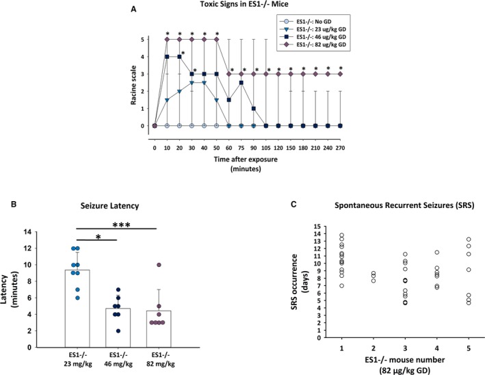
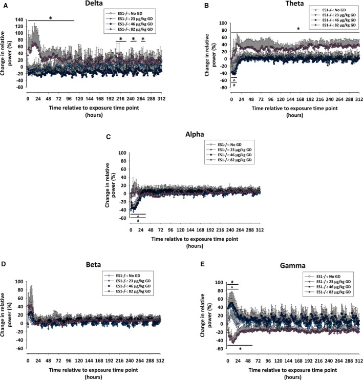
Electroencephalography (EEG) electrode-implanted ES1-/- and wild-type (C57BL/6) mice were exposed to various seizure-inducing doses of GD, treated with atropine sulfate and the oxime HI-6 at 1 minute after exposure, and administered midazolam at 15-30 minutes following the onset of seizure activity. The latency of acute seizure onset and spontaneous recurrent seizures (SRS) was assessed, as were changes in EEG power spectra. At 2 weeks after GD exposure, neurodegeneration and neuroinflammation were assessed.
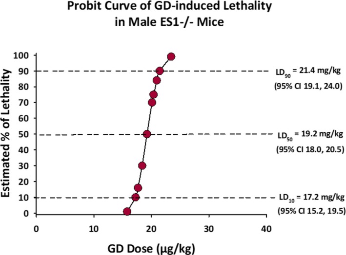
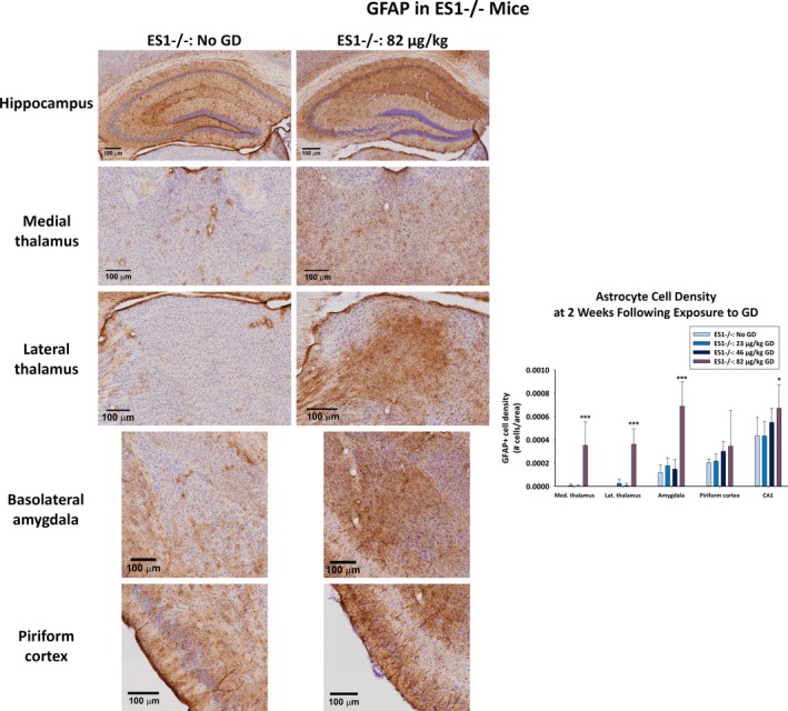
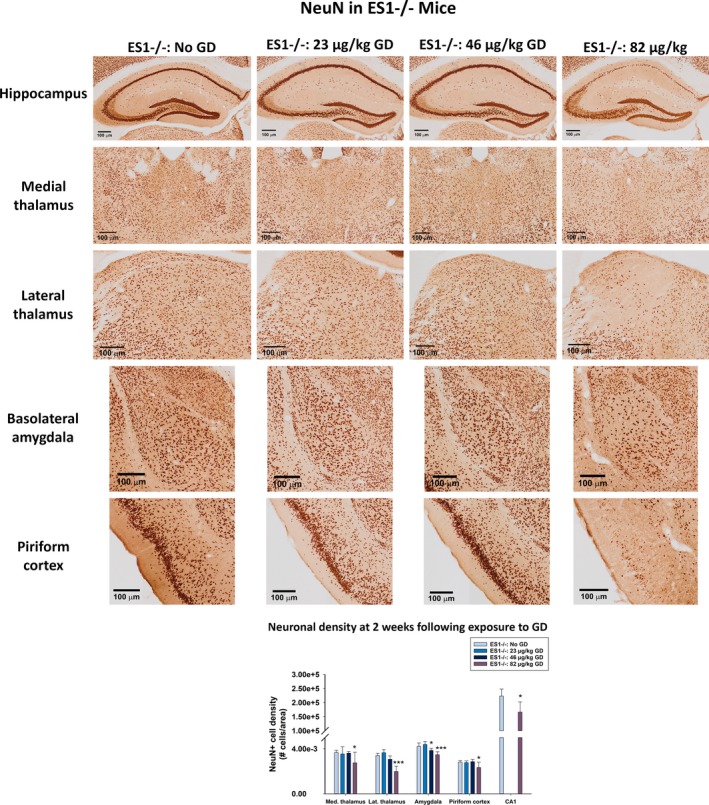
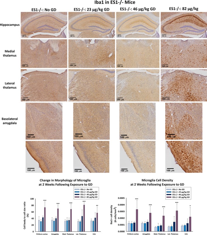
GD-exposed ES1-/- mice displayed a dose-dependent response in seizure severity. Only ES1-/- mice exposed to the highest tested dose of GD developed SE, subchronic alterations in EEG power spectra, and SRS. Degree of neuronal cell loss and neuroinflammation were dose-dependent; no significant neuropathology was observed in C57BL/6 mice or ES1-/- mice exposed to lower GD doses.
The US Food and Drug Administration (FDA) animal rule requires the use of relevant animal models for the advancement of MCMs against CWNAs. We present evidence that argues for the use of the ES1-/- mouse model to screen anticonvulsant, antiepileptic, and/or neuroprotective drugs against GD-induced toxicity, as well as to identify mechanisms of GD-induced epileptogenesis.
接触化学战神经毒剂(CWNAs),如沙林(GD),会导致癫痫持续状态(SE),如果治疗延迟,这种状态会对苯二氮䓬类药物产生抗药性,从而增加癫痫发生、严重神经病理学和长期行为认知缺陷的风险。啮齿动物模型广泛用于评估针对 CWNA 暴露的新型医疗对策(MCMs),通常表达血浆羧酸酯酶,这是一种参与某些有机磷化合物代谢的酶。为了更好地预测新型 MCMs 对人类伤亡 CWNA 暴露的疗效,使用反映人类状况的适当动物模型至关重要。我们描述了 GD 暴露后延迟抗惊厥治疗对血浆羧酸酯酶敲除(ES1-/-)小鼠致痫、致癫痫和神经病理学影响的综合特征。
植入脑电图(EEG)电极的 ES1-/-和野生型(C57BL/6)小鼠暴露于各种致痫剂量的 GD 中,在暴露后 1 分钟用硫酸阿托品和肟 HI-6 治疗,并在癫痫发作活动开始后 15-30 分钟给予咪达唑仑。评估急性发作开始和自发性复发性癫痫发作(SRS)的潜伏期,以及 EEG 功率谱的变化。GD 暴露后 2 周,评估神经退行性变和神经炎症。
GD 暴露的 ES1-/- 小鼠在癫痫严重程度上表现出剂量依赖性反应。只有暴露于最高测试剂量 GD 的 ES1-/- 小鼠才会出现 SE、亚慢性 EEG 功率谱改变和 SRS。神经元细胞丢失和神经炎症的程度呈剂量依赖性;在 C57BL/6 小鼠或暴露于较低 GD 剂量的 ES1-/- 小鼠中未观察到明显的神经病理学变化。
美国食品和药物管理局(FDA)动物规则要求使用相关动物模型来推进针对 CWNAs 的 MCMs。我们提供的证据表明,需要使用 ES1-/- 小鼠模型来筛选抗惊厥、抗癫痫和/或神经保护药物,以对抗 GD 诱导的毒性,以及识别 GD 诱导的癫痫发生机制。