Swiss Light Source, Paul Scherrer Institut, Villigen PSI, 5232, Switzerland.
Architecture et Réactivité de l'ARN, Université de Strasbourg, Institut de Biologie Moléculaire et Cellulaire du CNRS, Strasbourg, 67084, France.
RNA. 2019 Feb;25(2):173-192. doi: 10.1261/rna.068437.118. Epub 2018 Nov 8.
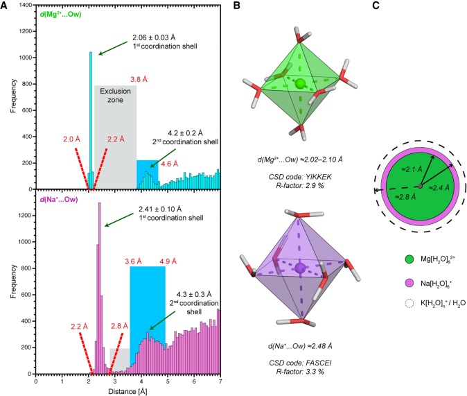
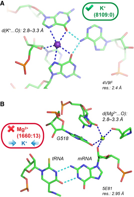
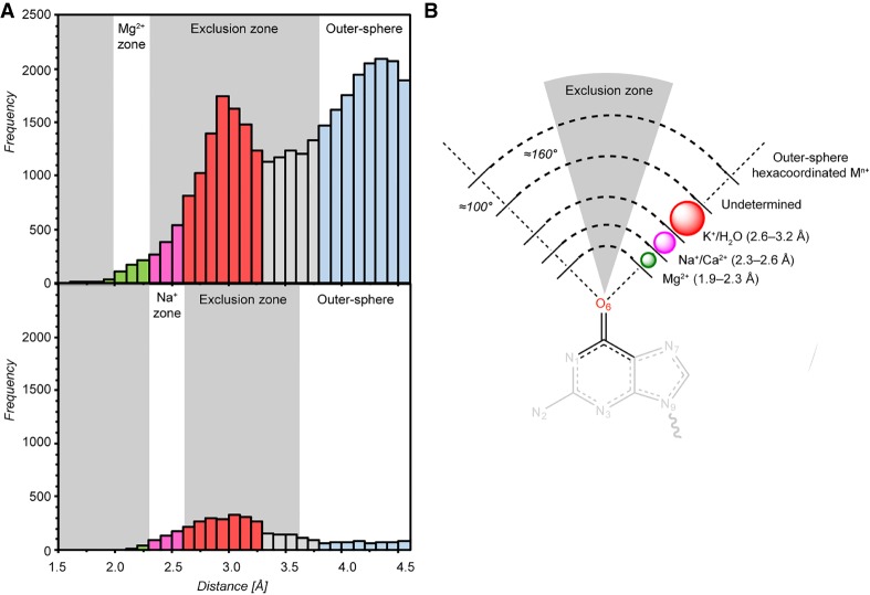
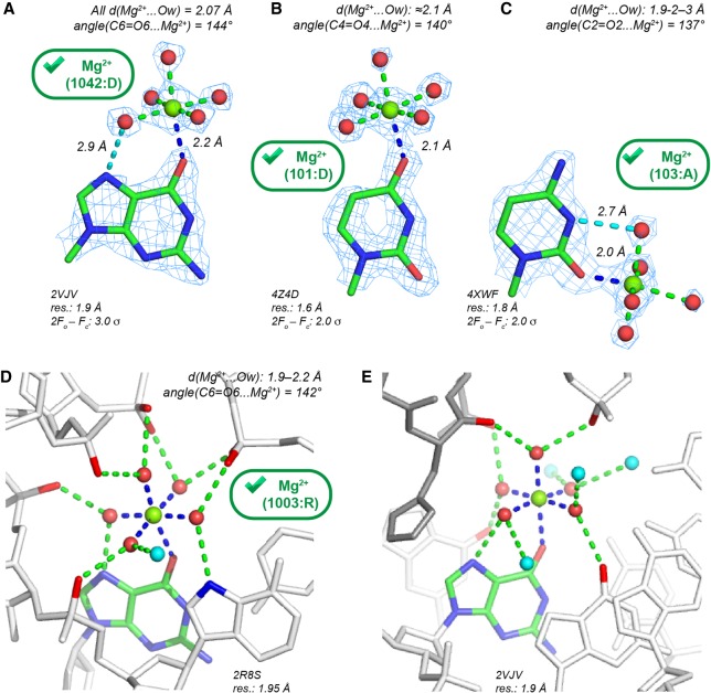
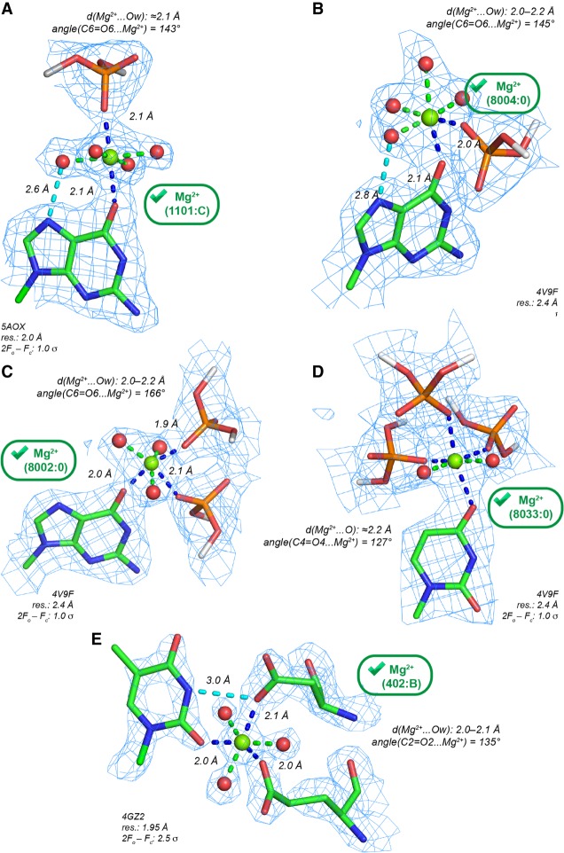
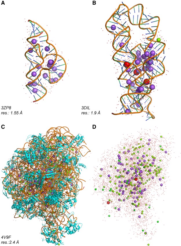
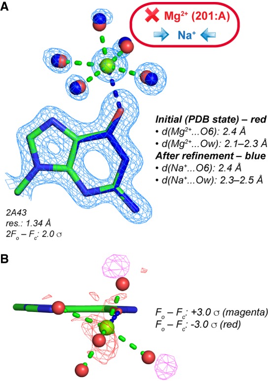
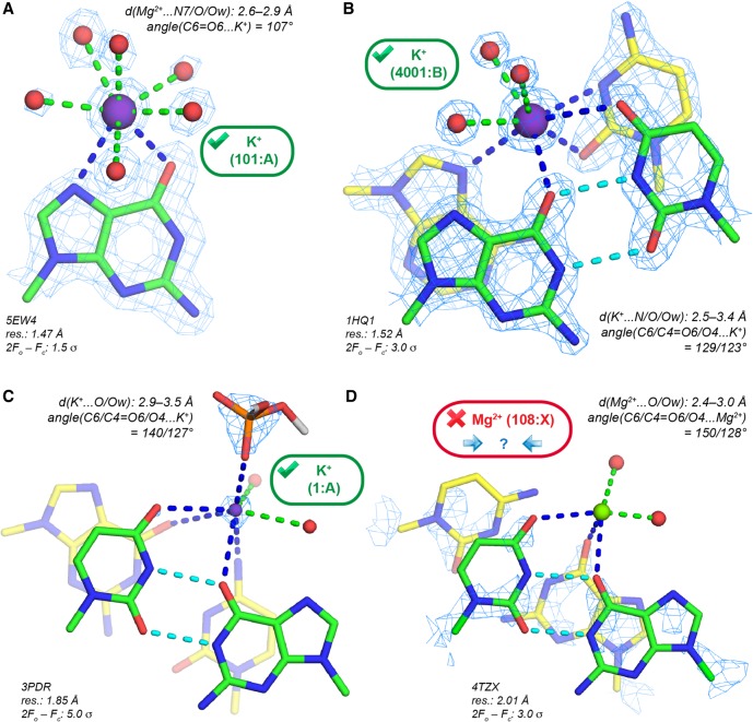
Precise knowledge of Mg inner-sphere binding site properties is vital for understanding the structure and function of nucleic acid systems. Unfortunately, the PDB, which represents the main source of Mg binding sites, contains a substantial number of assignment issues that blur our understanding of the functions of these ions. Here, following a previous study devoted to Mg binding to nucleobase nitrogens, we surveyed nucleic acid X-ray structures from the PDB with resolutions ≤2.9 Å to classify the Mg inner-sphere binding patterns to nucleotide carbonyl, ribose hydroxyl, cyclic ether, and phosphodiester oxygen atoms. From this classification, we derived a set of "prior-knowledge" nucleobase Mg binding sites. We report that crystallographic examples of trustworthy nucleobase Mg binding sites are fewer than expected since many of those are associated with misidentified Na or K We also emphasize that binding of Na and K to nucleic acids is much more frequent than anticipated. Overall, we provide evidence derived from X-ray structures that nucleobases are poor inner-sphere binders for Mg but good binders for monovalent ions. Based on strict stereochemical criteria, we propose an extended set of guidelines designed to help in the assignment and validation of ions directly contacting nucleobase and ribose atoms. These guidelines should help in the interpretation of X-ray and cryo-EM solvent density maps. When borderline Mg stereochemistry is observed, alternative placement of Na, K, or Ca must be considered. We also critically examine the use of lanthanides (Yb, Tb) as Mg substitutes in crystallography experiments.
准确了解 Mg 的内配位结合位点性质对于理解核酸系统的结构和功能至关重要。不幸的是,PDB 作为 Mg 结合位点的主要来源,存在大量的结构解析问题,这些问题模糊了我们对这些离子功能的理解。在这里,我们在前一项研究的基础上,对 PDB 中分辨率≤2.9 Å 的核酸 X 射线结构进行了调查,以对核苷酸羰基、核糖羟基、环醚和磷酸二酯氧原子的 Mg 内配位结合模式进行分类。通过这种分类,我们得出了一套“先验知识”的碱基 Mg 结合位点。我们报告说,可靠的碱基 Mg 结合位点的晶体学实例比预期的要少,因为其中许多与错误识别的 Na 或 K 有关。我们还强调,Na 和 K 与核酸的结合比预期的要频繁得多。总的来说,我们提供了源自 X 射线结构的证据,表明碱基是 Mg 的不良内配位结合体,但对单价离子具有良好的结合能力。根据严格的立体化学标准,我们提出了一套扩展的指南,旨在帮助直接与碱基和核糖原子接触的离子的分配和验证。这些指南应该有助于解释 X 射线和 cryo-EM 溶剂密度图。当观察到 Mg 立体化学的边界情况时,必须考虑 Na、K 或 Ca 的替代位置。我们还批判性地检查了镧系元素(Yb、Tb)在晶体学实验中作为 Mg 替代物的使用。