Department of Biochemistry, National Defense Medical Center, Taipei City, 114, Taiwan, Republic of China.
Division of Hematology/Oncology, Department of Medicine, Tri-Service General Hospital, National Defense Medical Center, Taipei City, 114, Taiwan, Republic of China.
J Biomed Sci. 2018 Nov 15;25(1):81. doi: 10.1186/s12929-018-0478-5.
Metformin is the most commonly used first-line medicine for type II diabetes mellitus. Acting via AMP-activated protein kinase, it has been used for more than 60 years and has an outstanding safety record. Metformin also offers protection against cancer, but its precise mechanisms remain unclear.
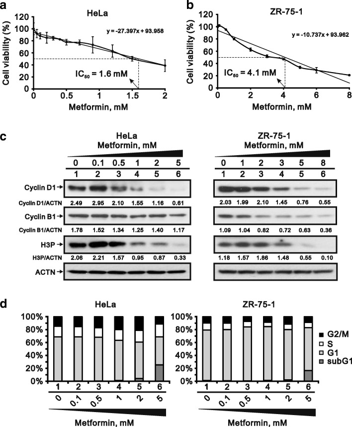
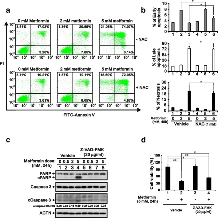
We first examined the cytotoxic effects of metformin in the HeLa human cervical carcinoma and ZR-75-1 breast cancer cell lines using assays of cell viability, cleaved poly-ADP-ribose polymerase, and Annexin V-fluorescein isothiocyanate apoptosis, as well as flow cytometric analyses of the cell cycle profile and reactive oxygen species (ROS). We later clarified the effect of metformin on p53 protein stability using transient transfection and cycloheximide chase analyses.
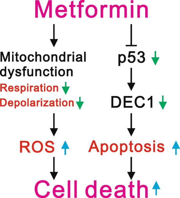
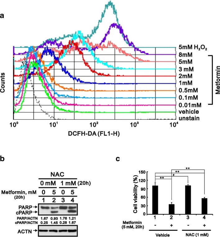
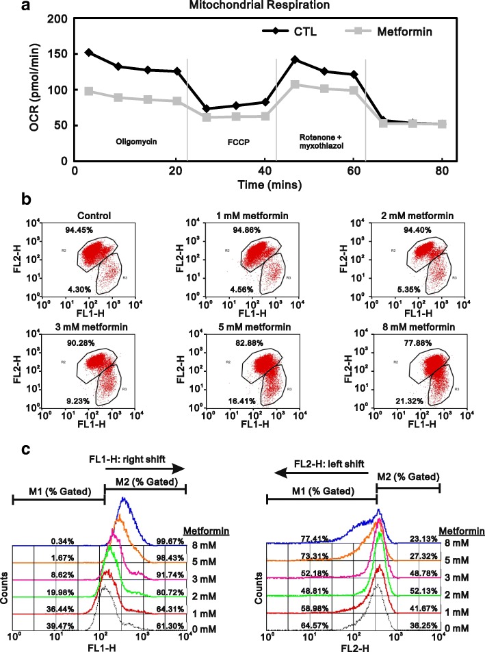
We observed that metformin represses cell cycle progression, thereby inducing subG1 populations, and had induced apoptosis through downregulation of p53 protein and a target gene, differentiated embryo chondrocyte 1 (DEC1). In addition, metformin increased intracellular ROS levels, but N-acetyl cysteine, a ROS scavenger, failed to suppress metformin-induced apoptosis. Further results showed that metformin disrupted the electron transport chain and collapsed the mitochondrial membrane potential, which may be the cause of the elevated ROS levels. Examination of the mechanisms underlying metformin-induced HeLa cell death revealed that reduced stability of p53 in metformin-treated cells leads to decreases in DEC1 and induction of apoptosis.
The involvement of DEC1 provides new insight into the positive or negative functional roles of p53 in the metformin-induced cytotoxicity in tumor cells.
二甲双胍是治疗 2 型糖尿病最常用的一线药物。通过激活 AMP 激活的蛋白激酶,它已经使用了 60 多年,并且具有出色的安全性记录。二甲双胍还能预防癌症,但确切的机制尚不清楚。
我们首先使用细胞活力测定、裂解多聚 ADP-核糖聚合酶和 Annexin V-异硫氰酸荧光素凋亡测定以及细胞周期谱和活性氧(ROS)的流式细胞分析,检测二甲双胍在 HeLa 人宫颈癌细胞和 ZR-75-1 乳腺癌细胞系中的细胞毒性作用。之后,我们使用瞬时转染和环己酰亚胺追踪分析阐明了二甲双胍对 p53 蛋白稳定性的影响。
我们观察到二甲双胍抑制细胞周期进程,从而诱导 subG1 群体,并通过下调 p53 蛋白和靶基因分化胚胎软骨细胞 1(DEC1)诱导细胞凋亡。此外,二甲双胍增加了细胞内 ROS 水平,但 ROS 清除剂 N-乙酰半胱氨酸未能抑制二甲双胍诱导的细胞凋亡。进一步的结果表明,二甲双胍破坏了电子传递链并使线粒体膜电位崩溃,这可能是 ROS 水平升高的原因。对二甲双胍诱导的 HeLa 细胞死亡机制的研究表明,二甲双胍处理细胞中 p53 稳定性降低导致 DEC1 减少并诱导细胞凋亡。
DEC1 的参与为 p53 在肿瘤细胞中对二甲双胍诱导的细胞毒性的正或负功能作用提供了新的见解。