1 Department of Pharmacology and Molecular Sciences, School of Medicine, Johns Hopkins University, Baltimore, MA, USA.
2 Center for Epigenetics, School of Medicine, Johns Hopkins University, Baltimore, MA, USA.
Mol Pain. 2018 Jan-Dec;14:1744806918817429. doi: 10.1177/1744806918817429. Epub 2018 Nov 19.
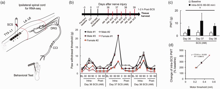
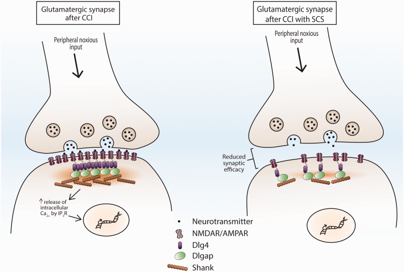
Spinal cord stimulation has become an important modality in pain treatment especially for neuropathic pain conditions refractory to pharmacotherapy. However, the molecular control of inhibitory and excitatory mechanisms observed after spinal cord stimulation are poorly understood. Here, we used RNA-seq to identify differences in the expression of genes and gene networks in spinal cord tissue from nerve-injured rats with and without repetitive conventional spinal cord stimulation treatment. Five weeks after chronic constrictive injury to the left sciatic nerve, male and female rats were randomized to receive repetitive spinal cord stimulation or no treatment. Rats receiving spinal cord stimulation underwent epidural placement of a miniature stimulating electrode and received seven sessions of spinal cord stimulation (50 Hz, 80% motor threshold, 0.2 ms, constant current bipolar stimulation, 120 min/session) over four consecutive days. Within 2 h after the last spinal cord stimulation treatment, the L4-L6 spinal segments ipsilateral to the side of nerve injury were harvested and used to generate libraries for RNA-seq. Our RNA-seq data suggest further increases of many existing upregulated immune responses in chronic constrictive injury rats after repetitive spinal cord stimulation, including transcription of cell surface receptors and activation of non-neuronal cells. We also demonstrate that repetitive spinal cord stimulation represses transcription of several key synaptic signaling genes that encode scaffold proteins in the post-synaptic density. Our transcriptional studies suggest a potential relationship between specific genes and the therapeutic effects observed in patients undergoing conventional spinal cord stimulation after nerve injury. Furthermore, our results may help identify new therapeutic targets for improving the efficacy of conventional spinal cord stimulation and other chronic pain treatments.
脊髓刺激已成为疼痛治疗的重要手段,尤其适用于对药物治疗有抗性的神经性疼痛病症。然而,脊髓刺激后观察到的抑制和兴奋机制的分子控制仍知之甚少。在这里,我们使用 RNA-seq 来鉴定神经损伤大鼠脊髓组织中基因和基因网络表达的差异,这些大鼠分为接受重复常规脊髓刺激治疗和未接受治疗的两组。在左侧坐骨神经慢性缩窄性损伤后 5 周,雄性和雌性大鼠被随机分为接受重复脊髓刺激治疗或不治疗两组。接受脊髓刺激治疗的大鼠接受硬膜外微型刺激电极放置,并接受为期 4 天的 7 次脊髓刺激(50 Hz,80%运动阈值,0.2 ms,恒流双极刺激,120 min/session)。在最后一次脊髓刺激治疗后 2 小时内,收获损伤侧 L4-L6 脊髓节段,用于生成 RNA-seq 文库。我们的 RNA-seq 数据表明,在重复脊髓刺激后,慢性缩窄性损伤大鼠的许多现有上调免疫反应进一步增加,包括细胞表面受体的转录和非神经元细胞的激活。我们还证明,重复脊髓刺激抑制了几个关键突触信号基因的转录,这些基因编码突触后密度中的支架蛋白。我们的转录研究表明,特定基因与神经损伤后接受常规脊髓刺激的患者观察到的治疗效果之间可能存在潜在关系。此外,我们的结果可能有助于确定改善常规脊髓刺激和其他慢性疼痛治疗效果的新治疗靶点。