Jiang Xin, Chen Dong
Department of Spine Surgery, China-Japan Friendship Hospital, Beijing, China,
J Pain Res. 2018 Oct 31;11:2687-2698. doi: 10.2147/JPR.S162988. eCollection 2018.
Currently, there is a lack of comparative research about different lumbar disc patterns in patients with work-related chronic low back pain (CLBP) based on magnetic resonance imaging (MRI) analysis. Therefore, this study, on different patterns of lumbar disc degeneration or herniation in patients with CLBP, is valuable. In this study, we retrospectively investigated lumbar degenerative changes in patients with CLBP by using MRI analysis.
Two hundred and eighty-three patients (110 women and 173 men) with work-related CLBP were enrolled and divided into four groups based on intervertebral disc morphology from MRI analysis, including normal discs (ND) group, degenerative discs (DD) group, bulging discs (BD) group, and herniated discs (HD) group. Demographic characteristics, occupational information, Visual Analog Scale (VAS) scores, and Oswestry Disability Index (ODI) scores were analyzed. Moreover, multiple parameters were investigated in the MRI analysis.
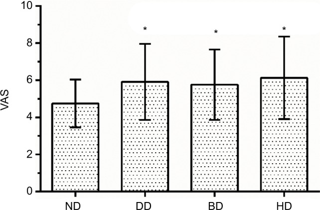
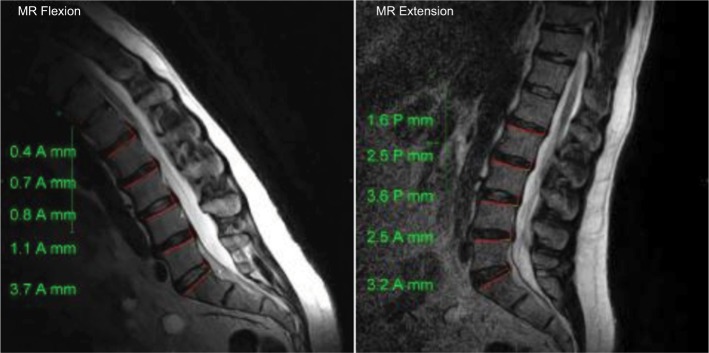
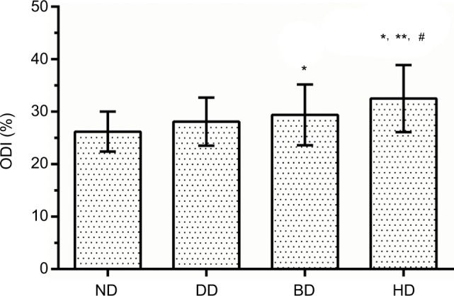
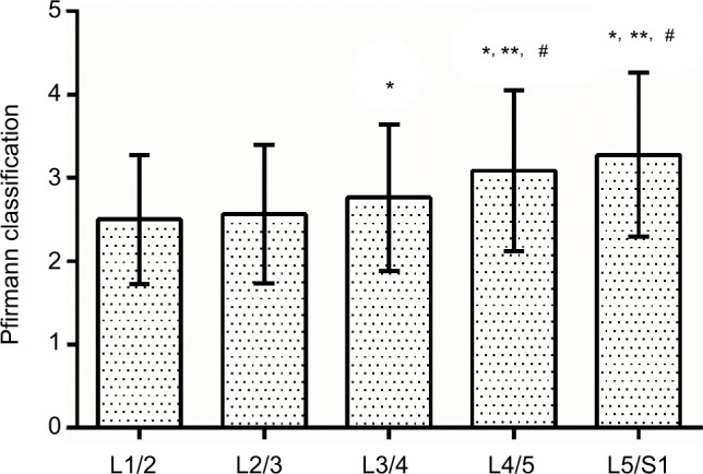
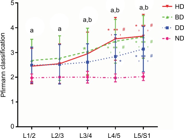
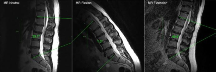
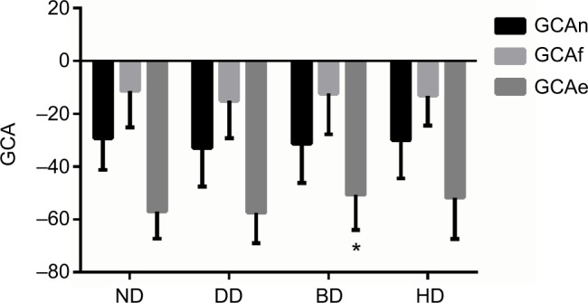
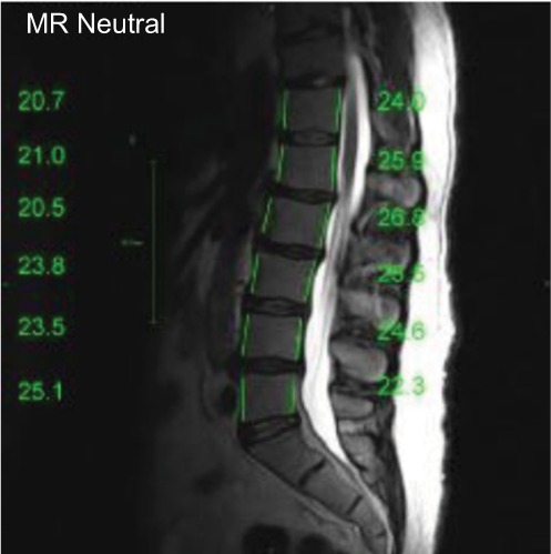
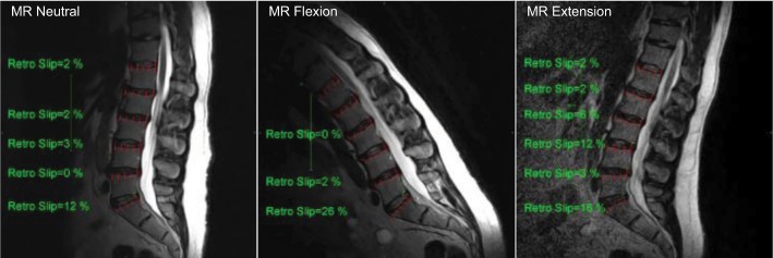
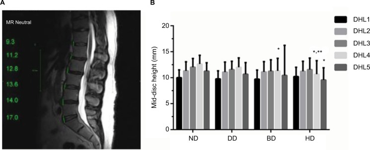
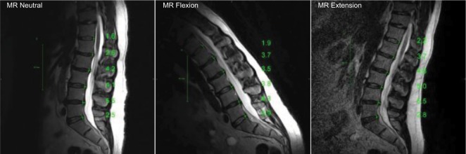
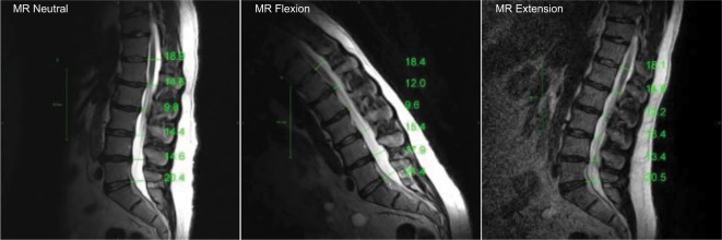
The mean age of all 283 patients was 41.8±12.0 years (range, 18-80) and the mean duration of CLBP for all patients was 24.5±24.9 months. There were no significant differences in the patients' BMI, history of smoking, and education level (>0.05). The three most common occupational types were manual worker, desk worker, and technician. The VAS and ODI scores of patients with CLBP in the DD, BD, and HD groups were significantly higher than those of patients in the ND group (<0.05). The degrees of degeneration of L4/5 and L5/S1 were significantly higher than those of other intervertebral discs (<0.05). The disc heights of L4/5 in the BD and HD groups were significantly lower than those of the ND group (<0.05) and the disc height of L5/S1 in the HD group was significantly lower than that of the ND group (<0.05). At the neutral position, the distances of L3/4, L4/5, and L5/S1 discs' bulge/herniation in the BD and HD groups were significantly higher than those in the ND and DD groups (<0.05).
In summary, more severe degenerative changes of lower lumbar discs (L4/5 and L5/S1) such as higher degree of degeneration of disc, lower disc height, and significant displacement of disc were found in patients with work-related CLBP based on MRI analysis.
目前,基于磁共振成像(MRI)分析,针对与工作相关的慢性下腰痛(CLBP)患者不同腰椎间盘模式的比较研究尚显不足。因此,本研究针对CLBP患者腰椎间盘退变或突出的不同模式具有重要价值。在本研究中,我们通过MRI分析回顾性研究了CLBP患者的腰椎退变情况。
纳入283例与工作相关的CLBP患者(110例女性和173例男性),并根据MRI分析的椎间盘形态将其分为四组,包括正常椎间盘(ND)组、退变椎间盘(DD)组、膨出椎间盘(BD)组和突出椎间盘(HD)组。分析了人口统计学特征、职业信息、视觉模拟评分(VAS)和Oswestry功能障碍指数(ODI)评分。此外,在MRI分析中还研究了多个参数。
283例患者的平均年龄为41.8±12.0岁(范围18 - 80岁),所有患者CLBP的平均病程为24.5±24.9个月。患者的体重指数、吸烟史和教育水平无显著差异(>0.05)。三种最常见的职业类型是体力劳动者、办公室职员和技术人员。DD组、BD组和HD组CLBP患者的VAS和ODI评分显著高于ND组患者(<0.05)。L4/5和L5/S1的退变程度显著高于其他椎间盘(<0.05)。BD组和HD组L4/5的椎间盘高度显著低于ND组(<0.05),HD组L5/S1的椎间盘高度显著低于ND组(<0.05)。在中立位时,BD组和HD组L3/4、L4/5和L5/S1椎间盘膨出/突出的距离显著高于ND组和DD组(<0.05)。
综上所述,基于MRI分析发现,与工作相关的CLBP患者下腰椎间盘(L4/5和L5/S1)存在更严重的退变改变,如椎间盘退变程度更高、椎间盘高度更低以及椎间盘明显移位。