• 文献检索
  • 文档翻译
  • 深度研究
  • 学术资讯
  • Suppr Zotero 插件Zotero 插件
  • 邀请有礼
  • 套餐&价格
  • 历史记录
应用&插件
Suppr Zotero 插件Zotero 插件浏览器插件Mac 客户端Windows 客户端微信小程序
定价
高级版会员购买积分包购买API积分包
服务
文献检索文档翻译深度研究API 文档MCP 服务
关于我们
关于 Suppr公司介绍联系我们用户协议隐私条款
关注我们

Suppr 超能文献

核心技术专利:CN118964589B侵权必究
粤ICP备2023148730 号-1Suppr @ 2026

文献检索

告别复杂PubMed语法,用中文像聊天一样搜索,搜遍4000万医学文献。AI智能推荐,让科研检索更轻松。

立即免费搜索

文件翻译

保留排版,准确专业,支持PDF/Word/PPT等文件格式,支持 12+语言互译。

免费翻译文档

深度研究

AI帮你快速写综述,25分钟生成高质量综述,智能提取关键信息,辅助科研写作。

立即免费体验

尸检系列中中耳解剖变异的内镜评估:204 耳分析。

Endoscopic evaluation of middle ear anatomic variations in autopsy series: analyses of 204 ears.

机构信息

Kocaeli Health Sciences University, Derince Training and Research Hospital, Department of Otorhinolaryngology - Head and Neck Surgery, Kocaeli, Turkey.

University of Istanbul, Istanbul Medical Faculty, Department of Otorhinolaryngology - Head and Neck Surgery, Istanbul, Turkey.

出版信息

Braz J Otorhinolaryngol. 2020 Jan-Feb;86(1):74-82. doi: 10.1016/j.bjorl.2018.10.002. Epub 2018 Nov 3.

DOI:10.1016/j.bjorl.2018.10.002
PMID:30472004
原文链接:https://pmc.ncbi.nlm.nih.gov/articles/PMC9422588/
Abstract

INTRODUCTION

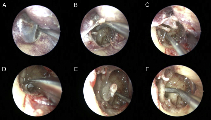
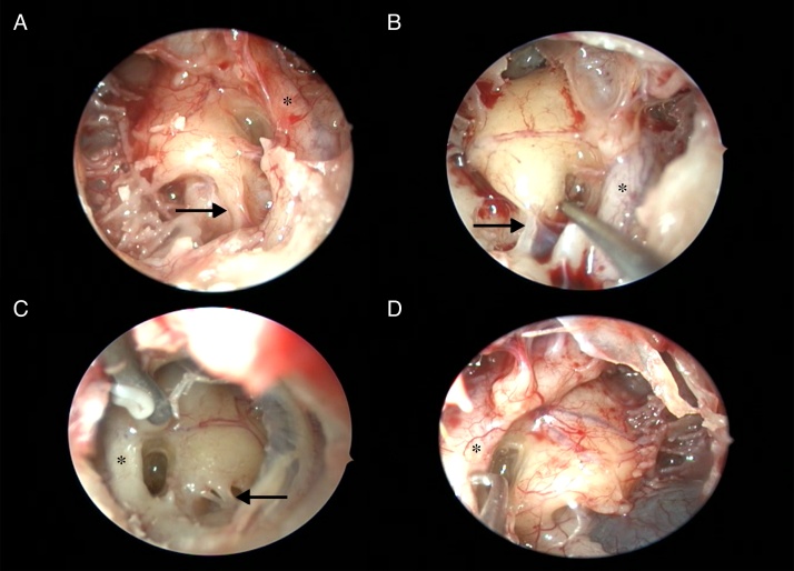
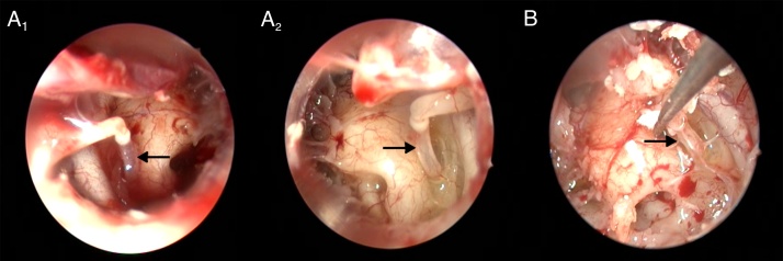
Microsurgery of the ear requires complete evaluation of middle ear surgical anatomy, especially the posterior tympanic cavity anatomy. Preoperative assessment of the middle ear cavity is limited by the permeability of eardrum and temporal bone density. Therefore, middle ear exploration is an extremely useful method to identify structural abnormalities and anatomical variations.

OBJECTIVE

The aim of this study is to determine anatomic variations of the middle ear in an autopsy series.

METHODS

All evaluations were performed in the Forensic Medicine Institute Morgue Department. The cases over 18 years of age, with no temporal bone trauma and history of otologic surgery included in this study.

RESULTS

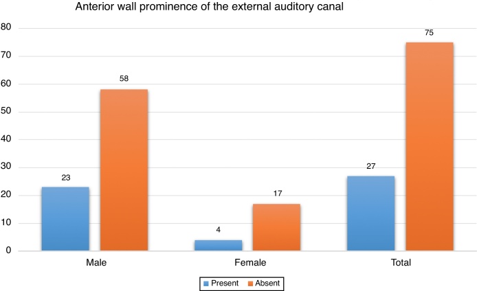
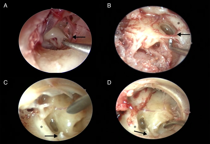
One hundred and two cadavers were included in the study. The mean age was 49.08±17.76 years. Anterior wall prominence of the external auditory canal was present in 27 of all cadavers (26.4%). The tympanic membrane was normal in 192 ears (94%) while several eardrum pathologies were detected in 12 ears (6%). Agenesis of the pyramidal eminence and stapedial tendon was found in 3 ears. While the ponticulus was bony ridge-shaped in 156 of 204 ears (76.4%), it was bridge-shaped in 25 ears (12.3%). The ponticulus was absent in 23 ears (11.3%). While complete subiculum was present in 136 of all ears (66.7%), incomplete subiculum was present in 21 ears (10.3%). Subiculum was absent in 47 ears (23%). Facial dehiscence was found in 32 ears and the round window niche was covered by a pseudomembrane in 85 ears (41.6%). A fixed footplate was present in 7.4% of all ears, and no persistent stapedial artery was seen in any cases.

CONCLUSION

The pseudomembrane frequency covering the round window niche was found different from reports in the literature. In addition, the frequency of the external auditory canal wall prominence has been reported for the first time.

摘要

介绍

耳部微创手术需要对中耳手术解剖结构进行全面评估,尤其是对后鼓室解剖结构的评估。中耳腔的术前评估受到鼓膜通透性和颞骨密度的限制。因此,中耳探查是一种非常有用的方法,可以识别结构异常和解剖变异。

目的

本研究旨在确定尸体解剖系列中耳的解剖变异。

方法

所有评估均在法医研究所太平间进行。本研究纳入年龄在 18 岁以上、无颞骨外伤和耳科手术史的病例。

结果

本研究纳入了 102 具尸体。平均年龄为 49.08±17.76 岁。所有尸体中,27 具(26.4%)存在外耳道前壁突出。192 只耳朵(94%)鼓膜正常,12 只耳朵(6%)发现多种鼓膜病变。3 只耳朵存在锥体隆起和镫骨肌腱发育不全。204 只耳朵中,156 只(76.4%)桥状鼓环,25 只(12.3%)为梁状鼓环,23 只(11.3%)鼓环缺失。所有耳朵中,136 只(66.7%)存在完整的鼓环下脚,21 只(10.3%)存在不完整的鼓环下脚,47 只(23%)鼓环下脚缺失。32 只耳朵面神经管存在骨裂,85 只耳朵(41.6%)圆窗龛被假性鼓膜覆盖。7.4%的耳朵存在固定的镫骨底板,所有病例均未见永存镫骨动脉。

结论

圆窗龛假性鼓膜覆盖的频率与文献报道不同。此外,首次报道了外耳道后壁突出的频率。

https://cdn.ncbi.nlm.nih.gov/pmc/blobs/c879/9422588/5b2e83ab0aa0/gr5.jpg
https://cdn.ncbi.nlm.nih.gov/pmc/blobs/c879/9422588/afebb87e1fa2/gr1.jpg
https://cdn.ncbi.nlm.nih.gov/pmc/blobs/c879/9422588/b2e1e6fe0ab4/gr2.jpg
https://cdn.ncbi.nlm.nih.gov/pmc/blobs/c879/9422588/910a67b4ff7f/gr3.jpg
https://cdn.ncbi.nlm.nih.gov/pmc/blobs/c879/9422588/abe3a01c596c/gr4.jpg
https://cdn.ncbi.nlm.nih.gov/pmc/blobs/c879/9422588/5b2e83ab0aa0/gr5.jpg

相似文献

1
Endoscopic evaluation of middle ear anatomic variations in autopsy series: analyses of 204 ears.尸检系列中中耳解剖变异的内镜评估:204 耳分析。
Braz J Otorhinolaryngol. 2020 Jan-Feb;86(1):74-82. doi: 10.1016/j.bjorl.2018.10.002. Epub 2018 Nov 3.
2
The variants of the retro- and hypotympanum: an endoscopic anatomical study.鼓室后隐窝及下鼓室的变异:一项内镜解剖学研究
Eur Arch Otorhinolaryngol. 2017 May;274(5):2141-2148. doi: 10.1007/s00405-017-4492-0. Epub 2017 Feb 27.
3
Otoendoscopy in cholesteatoma surgery of the middle ear: what benefits can be expected?耳内镜在中耳胆脂瘤手术中的应用:预期能带来哪些益处?
Otol Neurotol. 2008 Dec;29(8):1085-90. doi: 10.1097/MAO.0b013e318188e8d7.
4
Pyramidal eminence and subpyramidal space: an endoscopic anatomical study.锥体隆起和下锥体空间:内镜解剖研究。
Laryngoscope. 2010 Mar;120(3):557-64. doi: 10.1002/lary.20748.
5
Endoscopic anatomy of the posterior tympanum.鼓室后部的内镜解剖
Rev Laryngol Otol Rhinol (Bord). 2008;129(4-5):239-43.
6
Endoscopic classification of the external auditory canal for transcanal endoscopic ear surgery.经外耳道内镜耳部手术的外耳道内镜分类。
Eur Ann Otorhinolaryngol Head Neck Dis. 2019 Sep;136(4):247-250. doi: 10.1016/j.anorl.2019.03.005. Epub 2019 Mar 15.
7
[Congenital middle ear malformation: clinical analysis and discussion of classification].[先天性中耳畸形:临床分析与分类探讨]
Zhonghua Er Bi Yan Hou Tou Jing Wai Ke Za Zhi. 2019 Jul 7;54(7):481-488. doi: 10.3760/cma.j.issn.1673-0860.2019.07.001.
8
External auditory canal: Inferior, posterior-inferior, and anterior canal wall overhangs.外耳道:外耳道下壁、后下壁及前壁有骨质增生。
Int J Pediatr Otorhinolaryngol. 2018 Jun;109:138-143. doi: 10.1016/j.ijporl.2018.03.038. Epub 2018 Apr 4.
9
High-resolution computed tomographic study of the retrotympanum. Anatomic correlations.鼓室后隐窝的高分辨率计算机断层扫描研究。解剖学关联。
Surg Radiol Anat. 1998;20(3):215-20. doi: 10.1007/BF01628898.
10
Volume rendering of the tympanic cavity from micro-CT data.基于微计算机断层扫描(micro-CT)数据的鼓室容积再现
Folia Med Cracov. 2015;55(4):81-9.

引用本文的文献

1
Minimally invasive autopsy - endoscopic approach to post-mortem diagnostics.微创尸检——用于死后诊断的内镜方法。
Wideochir Inne Tech Maloinwazyjne. 2024 Mar;19(1):122-128. doi: 10.5114/wiitm.2023.134122. Epub 2023 Dec 29.
2
Videoautopsy-A Minimally Invasive Autopsy Method Using Endoscopic Techniques in Forensic Medicine: Clinical Features.视频尸检——法医学中一种使用内镜技术的微创尸检方法:临床特征
Diagnostics (Basel). 2024 Apr 24;14(9):884. doi: 10.3390/diagnostics14090884.
3
Application of Otoform to Study Variations of Sinus Tympani: A Novel Technique.

本文引用的文献

1
The variants of the retro- and hypotympanum: an endoscopic anatomical study.鼓室后隐窝及下鼓室的变异:一项内镜解剖学研究
Eur Arch Otorhinolaryngol. 2017 May;274(5):2141-2148. doi: 10.1007/s00405-017-4492-0. Epub 2017 Feb 27.
2
Radiological and audiometric evaluation of high jugular bulb and dehiscent high jugular bulb.高位颈静脉球及高位颈静脉球裂孔的放射学和听力评估
J Laryngol Otol. 2016 Nov;130(11):1059-1063. doi: 10.1017/S0022215116009166.
3
[Bone dehiscences in the middle ear in patients operated on ear diseases in the Otolaryngology Department of Jagiellonian University, Poland from 2008 to 2012].
使用耳模研究鼓窦隐窝变异:一种新技术
Indian J Otolaryngol Head Neck Surg. 2024 Feb;76(1):245-249. doi: 10.1007/s12070-023-04135-z. Epub 2023 Aug 20.
4
Juvenile Nasopharyngeal Angiofibroma in Adolescent Female with Subsequent ICA Canal Dehiscence: An Unknown Entity.青春期女性的青少年鼻咽血管纤维瘤伴随后续颈内动脉管裂开:一种未知的实体。
Indian J Otolaryngol Head Neck Surg. 2022 Dec;74(Suppl 3):4680-4687. doi: 10.1007/s12070-021-03008-7. Epub 2022 Jan 6.
5
Optical Coherence Tomography-Based Atlas of the Human Cochlear Hook Region.基于光学相干断层扫描技术的人类耳蜗钩区图谱。
J Clin Med. 2022 Dec 28;12(1):238. doi: 10.3390/jcm12010238.
6
The Endoscopic Anatomy of the External Acoustic Meatus and of the Middle Ear in Dry Temporal Bones: A Study Conducted Using Digital and Mobile Device Technology.干颞骨外耳道及中耳的内镜解剖:一项使用数字和移动设备技术开展的研究
Int Arch Otorhinolaryngol. 2022 Mar 8;26(4):e661-e665. doi: 10.1055/s-0041-1731813. eCollection 2022 Oct.
7
Landmarks for Proper Round Window Electrode Insertion in Cochlear Implantation.人工耳蜗植入中圆窗电极正确插入的标志。
J Int Adv Otol. 2022 May;18(3):210-213. doi: 10.5152/iao.2022.21435.
[2008年至2012年在波兰雅盖隆大学耳鼻喉科接受耳部疾病手术患者的中耳骨质缺损]
Otolaryngol Pol. 2014 May-Jun;68(3):124-8. doi: 10.1016/j.otpol.2013.11.001. Epub 2013 Nov 23.
4
The round window region and contiguous areas: endoscopic anatomy and surgical implications.圆窗区域及相邻区域:内镜解剖学及手术意义
Eur Arch Otorhinolaryngol. 2015 May;272(5):1103-12. doi: 10.1007/s00405-014-2923-8. Epub 2014 Feb 9.
5
High and dehiscent jugular bulb: clear and present danger during middle ear surgery.高位开放型颈静脉球:中耳手术中明确且现存的危险。
Surg Radiol Anat. 2014 May;36(4):369-74. doi: 10.1007/s00276-013-1196-z. Epub 2013 Sep 4.
6
Absence of both stapedius tendon and muscle.镫骨肌腱和肌肉均缺失。
Neurosciences (Riyadh). 2006 Apr;11(2):112-4.
7
A clinical and histopathologic study of jugular bulb abnormalities.颈静脉球异常的临床与组织病理学研究
Arch Otolaryngol Head Neck Surg. 2012 Jan;138(1):66-71. doi: 10.1001/archoto.2011.231.
8
Inferior retrotympanum revisited: an endoscopic anatomic study.经咽鼓管鼓室隐窝的内镜解剖研究
Laryngoscope. 2010 Sep;120(9):1880-6. doi: 10.1002/lary.20995.
9
A novel surgical technique for management of tinnitus due to high dehiscent jugular bulb.一种治疗高位颈静脉球高位开放引起的耳鸣的新手术技术。
Otolaryngol Head Neck Surg. 2010 Apr;142(4):576-81. doi: 10.1016/j.otohns.2009.12.007.
10
The recesses of the retro-tympanum.鼓室后隐窝
Rom J Morphol Embryol. 2010;51(1):61-8.